Welcome

I struggled to find PEM resources for my CT3 year, despite the variety of excellent resources out there. I hope this website will help point you in the right direction. I'm not a PEM expert, but am following the guidance CEM have issued (in the form of a syllabus) to put together this page. This page is not endorsed by CEM, and any mistakes are mine.

Please comment with corrections, additions and further suggestions.

All the information here is collected from the internet, and it might be out of date or inaccurate, so please use your judgement and adhere to your hospital's protocols. If you do notice any errors or omissions please comment so we can put them right!

To navigate, decide whether you want to start with a PMP or a PAP. You can then select which PMP or PAP you want to look at. You will then be taken to the summary page for that PMP, with links expanded topic collections. If you know what topic you want to look at already, click on the link on the right hand side.
Showing posts with label PAP1. Show all posts

Friday, 5 June 2015

Functional Abdominal Pain

Functional abdominal pain is mentioned in the higher syllabus for abdominal pain, and not the core syllabus. It is common in children, and some resources suggest it has a non-organic cause in most cases - some don't. Either way, a cause is difficult to elucidate, with a cause identified in <10%. It is important to consider physical and psychological factors. Functional abdominal syndrome is abdominal pain for 25% of the time, with other symptoms.
Apley's law: the further a recurrent abdominal pain is from the umbilicus, the more likely it is to be organic.

40% of 7 year olds have at least one episode of abdominal pain, with peaks in incidence at 5 and 10 years old.

Recurrent pain:  more common in girls than boys
                           more common in children whose parents have GI problems
                           obesity
                           Winter

3 of more episodes of abdominal pain in three months, that affects daily activities. 
Pain not associated with eating, loss of daily functioning, no other disorder

Causes
There is no clear idea what causes recurrent abdominal pain. The biophysical model of disease suggests it's a response to biological factors, family and school interactions, family environment and critical life events. 
It is thought, by Rome III, there are three main categories of functional abdominal pain:
Duodenal Ulcers
Abdominal Migraine
Irritable Bowel Syndrome

Duodenal Ulcers or Functional Dyspepsia: 
Consider in epigastric pain that causes night time waking. Treat by giving PPIs. Test for and treat H Pylori. If symptoms do not respond, then get an endoscopy - if the endoscopy is normal, consider functional dyspepsia.

Irritable Bowel Syndrome
Intestinal dysmotility. Family history is common, and the infection may follow a GI infection. You normally get abdominal pain that is worse before defecation - and relieved by defacation. It can be helpeful to say to children that sometimes the insides of the intestine become so sensitive that some children can feel the food going round the bends. 
Peppermint oil may be helpful. 
Avoiding sorbitol can be helpful, and increasing intake of oats and linseed can help. 

Abdominal Migraine
Abdominal migraine is associated with travel sickness. This may be associated with a headache, but in some children the abdominal pain predominates. The pain is normally midline associated with vomiting and pallor. There is normally a history of migraine. 
Pizotifen may be helpful. 

Management
Make sure you differentiate between serious and dangerous diagnoses. Serious is a disruption to schooling and life. Dangerous is life threatening. 

- Urine culture and microscopy
- FBC, ESR, CRP, LFTs, U&E, Coeliac
- Stool microsccopy 
- Abdominal USS to exclude gall stones and PUJ obstruction
- Pain and life event diary

Red Flags
Unexplained fever
Weight loss and poor growth
Joint problems, rashes
Vomiting
Pain causing waking, referred to back or shoulders
Urinary symptoms, perianal disease, PR blood 
Age under 5 

References
http://learning.bmj.com/learning/module-intro/functional-recurrent-abdominal-pain-children-assessment-management.html?moduleId=10017102&searchTerm=%E2%80%9Cabdominal%E2%80%9D&page=1&locale=en_GB

http://gut.bmj.com/content/45/suppl_2/II43.full

Wednesday, 3 June 2015

Foreign Bodies

Children often swallow things. The management pretty much depends on what they have swallowed, and whether they have symptoms or not. If the child is coughing, consider that they may have inhaled the foreign body instead - the inhaled FB can act as a ball-valve and air can enter but not leave.

The Object
Button batteries, objects >5mm and sharp, and razor blades are considered dangerous. Button batteries are toxic, and have a slow but deep action, and can also cause direct pressure necrosis. Their effects may be seen after they have been removed. Open safety pins might be dangerous - sharp objects have a 15 - 35% risk of perforation.

Imaging
1. X-ray if likely to be radio-opaque or "dangerous".  Request a neck and chest x-ray- an abdomen is not needed, and irradiates the gonads un-necessarily.
Look at the x-ray carefully- common points that get stuck are:
- C6 is cricopharyngeal sling and upper oesophageal sphincter
- 15% get stuck in the midoesophagus where the aortic arch and carina push on the oesophagus.
- 15% get stuck in the lower oesophageal sphincter / oesophagogastric junction
Check carefully it is a coin and not a button battery. If you are not sure if the coin is in the oesophagus or not, do a lateral film. If the coin is in the oesophagus, it appears coronal. Tracheal objects appear in a sagittal orientation.

2. Dangerous Object - refer to surgeons where ever the object is. There is some debate about button batteries if they are below the diaphragm. Above the diaphragm - in the oesophagus, nose or throat, they need to come out ASAP.

3. Above diaphragm, symptomatic, - refer to the surgeons. Most (75%) objects impact in the upper oesophagus.
b. Mild or no symptoms - home, repeat x-ray 24 hours.

4. Below diaphragm
Reassure, return if symptoms develop

5. Asymptomatic, not seen - reassure and return if symptoms develop.
b. Symptomatic - refer to surgeons

There is no need to search the poo. It might take six weeks for the foreign body to come out.

Metal Detector
The metal detector can help prove whether a FB is above the diaphragm or not. They can confirm whether the coin has reached the stomach.

(flowchart from the amazing and well worth buying http://www.amazon.co.uk/Emergency-Care-Minor-Trauma-Children/dp/144412014X


References
http://dontforgetthebubbles.com/the-magic-coin/
http://dontforgetthebubbles.com/podcast-week-button-batteries/
http://lifeinthefastlane.com/ccc/inhaled-foreign-body/
http://lifeinthefastlane.com/top-ten-foreign-bodies/
http://wikem.org/wiki/Esophageal_foreign_body
http://www.annemergmed.com/article/S0196-0644(84)80573-9/abstract
http://lifeinthefastlane.com/paediatric-quiz-017/
http://emedicine.medscape.com/article/801821-treatment
http://www.ncbi.nlm.nih.gov/pubmed/15913481
http://learnpediatrics.com/body-systems/gastrointestinal/suspected-foreign-body-ingestion/
http://www.sciencedirect.com/science/article/pii/S0165587612006519
http://blog.clinicalmonster.com/2015/03/foreign-body-ingestions-in-children-by-abi-iyanone/
http://pediatriceducation.org/2005/03/28/
http://www.sinaiem.org/pearls/2015/05/26/fool-me-once/
http://www.amazon.co.uk/Emergency-Care-Minor-Trauma-Children/dp/144412014X
http://www.amazon.co.uk/books/dp/0199589569

Tuesday, 2 June 2015

Constipation

Constipation is common. DFTB has written some excellent pieces of work on this - I've written myself more of a summary using their resources, and some I've found from elsewhere. 

Definitions
RomeIII Criteria: 
≤2 stools per week for a duration of 2 months if patient >2 years
                               for duration of 4 months if patient <2 years
or with evidence of overflow incontinence (no stool, then diarrhoea, then no stool, etc), 
or stools that clog toilet

Chronic: >8 weeks
Happens in 5 - 30%of the child population, progressing to chronic in > 1/3 of patients

<3 months, 2-3 stools/ day, 8.5 hours mouth to rectum time
<2 years, <2/ day, 16 hours mouth to rectum time

Reservoir Constipation
- Too busy to poo, scared to poo leads to reservoir constipation. Stools get larger and harder. Pass a large diameter stool every 1-2 weeks. It's painful to pass these.
The rectum stretches. The internal sphincter struggles leading to a numb, toneless rectum. Chronically, can lead to anal fissures, which are painful so poo-ing is avoided. The stool continues to become harder and more painful to pass.

Hirschprung's Disease
1/500 live births
Normally diagnosed in newborns. 
Get abdominal distension that is relieved by rectal stimulation, or enemas.

Cow's Milk: Tolerance may lead to constipation - should be investigated by a specialist before avoiding cow's milk. 

Examination
Weigh the child
Abdo exam
Perianal exam – appearance, position, patency, fissures
Scoliosis + Gait 
Skin overlying the spine – discoloured/sinus/hairy patch/central pit
Gluteal muscles – is there asymmetry?
Neuro
No PR

Red Flag Features
Constipation from early infancy
Delay in meconium >48hrs
Ribbon stools
Abdo distension & vomiting
Abnormal appearance of anus including multiple anal fissures
Asymmetry/flattening of gluteals
Sacral agenesis, skin changes over spine
Skin changes overlying spine
Deformity of lower limbs – talipes
Abnormal neuromuscular signs

Treatment
This is summarised so clearly on CYP that I haven't re-written it
- Get rid of old, dark, hard and smelly poo
- Continue treatment for 3 months. 
- Make going to the toilet fun


References

Wednesday, 13 May 2015

Ovarian Torsion

Strangulated Hernias could rarely include an ovary - 4.9 cases/  100,000
Ovarian torsion occurs in young women (63% in 7-10yr old), and post-menopausal women
20% of cases occur during pregnancy

- Bowel takes a long time, so if there's a hard lump, think of a trapped ovary
- It might feel mobile like a bean
- Don't squish the ovary
- More commonly on the right hand side
- More likely if there's a cyst
- 60% of patients also have vomiting
- 3% have peritoneal signs
- 30% have no tenderness to palpation

Treatment
Ovaries can be difficult to reduce but don't become ischaemic as quickly as testicles.

References
http://pedemmorsels.com/ovarian-torsion/?utm_source=feedburner&utm_medium=twitter&utm_campaign=Feed%3A+FOAMEM+%28FOAM+RSS%29
http://www.rcemlearning.co.uk/modules/tummy-painagain/
http://radiopaedia.org/articles/ovarian-torsion
http://emlyceum.com/2012/06/21/ovarian-torsion-answers/

Friday, 8 May 2015

Paediatric Hernias

Epidemiology
Often present within 1st year of life.

Pathophysiology
Indirect - most hernias are indirect and extend through the internal and external rings. Often on the right hand side. 

Signs & Symptoms
- Asymptomatic bulge in the groin or scrotum - above the testicle
- May resolve when calm and supine
- Analgesia needed before reduction
- Gentle traction on the scrotum to help align the hernia sac with the external ring.
- While keeping gentle traction, squeeze distal to proximal 
- Apply pressure laterally with the index and thumb along each side of the hernia neck and inguinal canal.
- Imagine you are trying to stretch open the rings.
- Gently add more pressure distally and help reduce the hernia.
- This can take up to 40minutes

Incarceration or Strangulation
- Happens in 7 - 30% 
- Severe pain, bilious emesis, blood in stool, signs of peritonitis, redness and oedema on affected side of scrotum
- Don't attempt to manually reduce

References

Penile Problems

Penile problems in children


Hypospadius
Correct before 2 years of age. Do NOT circumcise as may need reconstructive surgery

Phimosis
- At 1 year old 50% boys have non-retractile foreskin, by 4yrs 10% and by 16yrs only 1%
- Non-retractile foreskin = balooning on micturition
- Topical corticosteroids can help.
- Encourage patients to maintain good hygiene and gently stretch the foreskin.

Emergency if causes acute urinary retention

Paraphimosis
Foreskin gets stuck in the retracted position.
Look for a hair tourniquet.
May be secondary to masturbation
Needs analgesia to reduce

Balanoposthitis
Balanitis = cellulitis of the glans
Posthesis = cellulitis of the foreskin

Can be irritant, bacterial or fungal.

Management
Needs warm baths
Rule out diabetes
Clotrimazole or antibiotic ointment
Normally get oral amox or trimeth too


References
http://emupdates.com/2009/04/01/900-balanitis-vs-balanoposthitis-causes-of-balanoposthitis-rx-conditions-prevented-by-circumcision/
http://www.wikem.org/wiki/Phimosis
http://emupdates.com/2009/04/01/899-management-of-phimosis-paraphimosis-causes-consequences-treatment/ 
http://www.bmj.com/content/346/bmj.f3678?sso= 
http://www.emrap.tv/index.php?option=com_content&view=article&id=106
http://www.wikem.org/wiki/Balanoposthitis
http://www.amazon.co.uk/Illustrated-Textbook-Paediatrics-STUDENTCONSULT-Online/dp/0723435650

Non Torsion Scrotum

Idiopathic Scrotal Oedema
Pathophysiology
"Cellulitis of the scrotum"
- Unknown cause
- Hypothesized that it represents a hypersensitivity reaction, similar to angioneurotic oedema

Signs
- May have a small scrotal scratch or insect bite 
- Pre-schoolers
- Starts as a small spot, then extends to cover half of the scrotum.
- No testicular tenderness - examine through unaffected skin

Management
- Self resolving
- NSAIDs and antibiotics have been used. 


Epididymo-orchitis
Pathophysiology
- Affects very young or very old.

Management
- Infants - exclude urinary tract abnormalities - renal USS + urine culture
- Post pubertal - consider sexual contact

Hydrocele of the cord
This often presents as a “third ball”. For elective management. 


Undescended Testes
4% incidence at birth (higher in premature babies), falling to 1% at age 1. 

Varicocoele
Thought of as “varicose veins” of the testicular veins.
More common at puberty

Signs
Classically feels like a “bag of worms”.
More commonly on the left, as testicular vein drains into higher-pressured left renal vein with a 90 degree turn 
Dullness/ heaviness / scrotal discomfort
Varices more prominent with standing or Valsalva
Does not trans-illuminate

Management
Need to exclude any other causes of obstruction at this level (e.g. renal tumour, renal vein thrombosis) --> especially if happens suddenly 
Treat surgically for symptomatic relief

Hydrocele
Happens if there is a patent processus vaginalis. 

Signs
Often asymptomatic bilateral scrotal swellings
Sometimes have a blueish discoloration
Transilluminate

Management
Most resolve spontaneously 
Surgery if persist beyond 18 - 24 months
If acute, check no inflammatory process 

References
And references on testicular torsion page

Testicular Torsion

Anatomy

The spermatic cord starts at the deep inguinal ring, then enters the scrotum at the superficial inguinal ring.
The testicular appendage, of hydatid of Morgagni, is a remnant of the Mullerian duct in 90% of cases. It can become twisted, mimicking symptoms of testicular torsion.

Pathophysiology
The testicle can twist and tighten up around the spermatic cord. The twist can happen outside of the scrotum, normally at the external inguinal ring, or inside of the scrotum. Inside is more common, especially in adolescents and young adults, especially if there is a bell clapper deformity.


In 5-8% of cases, this twisting is triggered by mild or moderate trauma.

Epidemiology
Occurs soon after birth, or at puberty.
Incidence in males <25 is 1 in 4000

Although it has no physiological function, it can be medically significant in that it can, occasionally, undergo torsion (i.e. become twisted), causing acute one-sided testicular pain and may require surgical excision to achieve relief. 1/3 of patients present with a palpable "blue dot" discoloration on the scrotum. This is nearly diagnostic of this condition. Although if clinical suspicion is high for testicular torsion, a surgical exploration of the scrotum is warranted.

Symptoms
Sudden onset, severe testicular pain - in 2/3 of the canal
Half of all torsions start in the night time
Pain not relieved by elevation of the scrotum
Swelling of the testis or scrotum, oedema or erythema of scrotal skin
Pain may be referred to abdominal or inguino-scrotal regions
No fever or urethral discharge

Signs
High riding testicle with horizontal lie
Loss of cremesteric reflex - Prohn's sign
Large testicle
Blue dot sign - more indicative of testicular appendage torsion

Undescended testes are at higher risk - empty scrotum, painful lump in the groin

Intermittent Testicular Torsion
May get spontaneous de-torsion. Short periods of groin pain + vomiting, then spontaneous relief.


TWIST Score

Proposed score for assessing testicular torsion in children
Finding                          Points
Testicular swelling          2
Hard testicle                  2
Absent cremasteric reflex  1
Nausea or vomiting          1
High-riding testicle          1

Treatment
Analgesia
Urgent Exploration
Consider manual detorsion if urologist not available - "open the book" - twist outward and laterally. May need to be done 2-3x for complete pain relief.


References
http://wikem.org/wiki/Testicular_torsion
http://radiopaedia.org/articles/testicular-torsion
http://emj.bmj.com/content/17/5/381.full
http://www.bmj.com/content/341/bmj.c3213?sso=
http://www.emrap.org/episode/2011/december/testicular
http://wikem.org/wiki/Testicular_Torsion
http://dontforgetthebubbles.com/testicular-trouble/
http://academiclifeinem.com/patwari-academy-videos-testicular-torsion-and-acute-scrotal-pain/
http://dontforgetthebubbles.com/saving-balls-101-inguinoscrotal-masses/
http://learning.bmj.com/learning/module-intro/testicular-torsion.html?moduleId=10029430&searchTerm=%E2%80%9Ctorsion%E2%80%9D&page=1&locale=en_GB
http://learning.bmj.com/learning/module-intro/scrotal-swellings-diagnosis-management.html?moduleId=5003328&searchTerm=%E2%80%9Ctorsion%E2%80%9D&page=1&locale=en_GB
http://www.enlightenme.org/learning-zone/tummy-pain%E2%80%A6again

http://dontforgetthebubbles.com/saving-balls-101-acute-scrotum/

Thursday, 1 May 2014

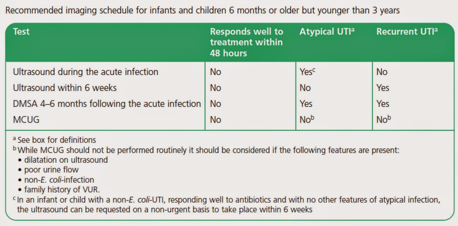
Urinary Tract Infections in Children

Up to 7% of girls and 2% of boys will have a symptomatic, culture confirmed urinary tract infection (UTI) by the age of six. The prevalence of UTI in febrile infants is approximately 7%. After a UTI, 10-30% of children have scarring


Clinical Features

Infants and children presenting with an unexplained fever of 38°C or higher, for over 24 hours, should have a urine sample tested. Infants and children with an alternative site of infection should not have a urine sample tested. When infants and children with an alternative site of infection remain unwell, urine testing should be considered after 24 hours at the latest.

How to test
Urgent microscopy and culture <3years old
>3 or no microscopy, dipstick testing may act as a substitute
Obtaining a Sample
Clean catch - less likely to be contaminated
Urine bags - useful to rule out, but not to rule in
Urine collection pads
Cotton wool balls, gauze and sanitary towels should not be used to collect urine in infants and children.

Treatments
Upper urinary tract infection - oral antibiotics (cephalosporin or co-amoxiclav) for 7 - 10  days
Lower urinary tract infections - oral antibiotics for 3 days.

Follow Up


References
http://learning.bmj.com/learning/module-intro/urinary-tract-infection-children-diagnosis-treatment-long-term-management-.html?moduleId=10011035&searchTerm=%E2%80%9CUTI%20children%E2%80%9D&page=1&locale=en_GB

http://www.enlightenme.org/the-learning-zone/node/4310
http://guidance.nice.org.uk/CG54/QuickRefGuide/pdf/English
http://dontforgetthebubbles.com/6-pem-papers-change-practice-6-getting-urine-neonates/
http://bestbets.org/bets/bet.php?id=34

Saturday, 29 March 2014

Mesenteric Adenitis

Mesenteric adenitis is a difficult diagnosis to make, but it is always a diagnosis of exclusion. There isn't much FOAMEd that I can find on mesenteric adenitis, and the trusty sunflower book doesn't actually say a lot about it. It lumps mesenteric adenitis with non-specific abdominal pain, and says NSAP is less severe than appendicitis, often accompanied by an URTI with cervical lymphadenopathy.

Pathophysiology
- Self-limiting illness
- Often affects the lymph nodes in the right lower quadrant only

- Pathogens are thought multiply in mesenteric lymph nodes. On gross pathology, lymph nodes are enlarged and soft. On microscopy, there is non-specific hyperplasia and when suppurative, there is necrosis and pus.

Mesenteric adenitis has a number of causes:
    variety of viruses
    Yersinia enterocolitica (Europe, North America and Australia)
    Helicobacter jejuni
    Campylobacter jejuni
    Salmonella spp
    Shigella spp

Treatment and prognosis
Self-limiting, and typically abates over the course of a few weeks.

Interestingly, when mesenteric adenitis (or appendicitis) occurs in childhood or adolescence, there is a significantly reduced risk of ulcerative colitis later in life.

Differential diagnosis
The clinical differential includes:
    acute appendicitis
        lymph nodes are generally smaller and fewer
        appendix is abnormal
    intussusception
    Meckel's diverticulitis

http://radiopaedia.org/articles/mesenteric-adenitis

Monday, 24 March 2014

Intussusception

Intussusception is a diagnosis I always worry about missing. I've seen it a few times - thankfully when I've been in a centre with paediatric surgery on site. There doesn't seem to be a big tip about how to diagnose it - except to always think of it as a differential. The few cases I've seen have presented with inconsolable abdominal pain with lots and lots of crying - and that has been the predominant symptom.

Epidemiology
- Happens mostly between 3months to 6 years
- Most common 3-12 months but can happen in elderly and adult patients
- 60% of cases are <1 year, 90% <2 years
- Boys: girls 3:2

Patho-physiology

A part of the bowel is pulled into the distal lumen, and peristalsed forward. The bit that is pulled (the prolapsed part) is called the intussusceptum. The bit of bowel that receives the intussusceptum is called the intussuscipiens. This process is likened to a piece of bowel telescoping in on itself.
Venous return gets compromised causing swelling, and restricted blood flow. Eventually arterial supply to the bowel is interrupted and ischaemia and necrosis happens.

This can occur anywhere:
ileocolic - most common (75-95%)
ileoileocolic - second most common
There is often a "lead point" that causes the intusussception - maybe a hypertrophied Peyer’s Patch. In adults a lesion is more likely. It is a complication of HSP in 2-6% of cases - generally in children >2yrs of age. HSP related intussusception is more likely to be ileocolic.

Clinical Features
- Episodes of abdominal pain - often 15- 20 minutes apart.
- Frequency and severity increase as intestinal oedema increases.
- Vomiting happens in some children. It may be billious.
- There is blood in the stools. In 75% of people with non visible blood stools, occult blood is positve. Jelly stools only happen in 50% of cases.
- Diarrhoea
- The classic triad of colicky abdominal pain, vomiting and redcurrent jelly stools - in 21% of cases only

InvestigationsAbdominal x-rays may demonstrate a soft tissue mass (typically in the right upper quadrant in children) with a bowel obstruction proximal to it. They are negative in 20% of cases! Some suggest if there is no air in the caecum, intusussception is more likely.

Abdominal ultrasound in comparison has 98–100% sensitivity and 88% specificity with a negative predictive value of 100%

Treatment
Air enema
More likely to need operative intervention if:
  Intussusception present >48 hours
  Age <3 months
  Age >5 years (higher likelihood of pathologic lead point)
Even after successful nonoperative reduction the recurrence risk is 10%.

http://pedemmorsels.com/intussusception/
http://www.bestbets.org/bets/bet.php?id=00388
http://bestbets.org/bets/bet.php?id=2372
http://empem.org/2011/09/intussusception/
http://pedemmorsels.com/hsp-and-intussusception/
http://www.pemcincinnati.com/blog/briefs-thoughts-about-abdominal-x-rays-in-intussusception/
http://emj.bmj.com/content/12/3/182.abstract
http://empem.org/2011/10/intussusception-rotavirus-vaccine-risk/
http://westjem.com/images/intussusception-status-post-roux-en-y-gastric-bypass.html
http://radiopaedia.org/articles/intussusception
http://pedemmorsels.com/intussusception/
http://www.pemcincinnati.com/blog/intussusception-1/
http://www.pemcincinnati.com/blog/intussusception-2/
http://www.pemcincinnati.com/blog/intussusception-3/
http://emupdates.com/2009/04/01/intussusception-ssx-dx-rx/
https://www.emrap.org/episode/2008/october/pediatric

Friday, 21 March 2014

Appendicitis

Appendicitis presents similarly in children and in adults, so it is difficult to separate the two age groups out entirely...so I'm not going to try too hard to do so! The important thing to remember is that atypical signs can lead to late presentations in children (and the elderly).

Pathophysiology


Initially the visceral pain is felt. This is vague, colicky, and in the midline. The appendix is embryologically a mid-gut organ, so pain in the appendix activates sympathetic fibres which enter the spinal cord at T10. This is why the pain is referred to the peri-umbilical area.

Later, somatic pain is felt. This is more constant, and more local. The peritoneum is innervated by the intercostal nerves.

Clinical Features
The Alvardo score can be useful for scoring appendicitis to see whether you think this is low risk. I've rarely seen this used in clinical practice but occasionally use it if the surgical SHO is being difficult and requesting bloods for a barn door appendix. 

M=Migration of pain to RIF             (1)
  (McBurney’s point which lies 1/3 of the way along an imaginary line from the anterior superior iliac spine to the umbilicus and indicates where the inflamed appendix normally lies.)
A=Anorexia                                     (1)
N=Nausea and vomiting                   (1)
T=Tenderness in RIF                        (2)
R=Rebound pain                              (1)
E=Elevated temperature                   (1)
L=Leukocytosis                               (2)
S=shift of WBC to left                      (1)
TOTAL                                          /10
 
 
All factors are quite useful. Anorexia is present in 80% of patients with anorexia.

Rovsing’s sign- press deeply in the LIF for 30s, release suddenly, patient experiences rebound tenderness in RIF.
Psoas sign- ask patient to lift flexed thigh against your hand placed just above the knee, patient experiences pain in RIF
PR: recent evidence suggests a painful PR exam is of little benefit with positive and negative predictive values of 0.44 and 0.54 respectively. Thankfully even the surgeons don't often ask for PRs in kids!


Investigations
Up to 30% of patients with appendicitis will have an abnormal urinalysis.
Plain abdominal x-ray has no role.  
USS has accuracy of about 90% (sensitivity 84% and specificity 88%)
CT has a greater overall accuracy of 94% (sensitivity 94%, specificity 95%) 
In children, USS is likely to be as good as CT.


Abdominal Pain in Children


As always, the syllabus is fairly vague. There are three groups of causes of abdominal pain in children (as I see it). 1. More likely in children, 2. more likely in adults but could happen in children, 3. referred or non- abdominal.

More Likely in Children
Testicular Torsion
Referred Scrotal Pain
Foreign body ingestion
Hernias

More Likely in Adults
Gall stones
Porphyria
Inflammatory bowel disease (Crohns) with or without toxic megacolon

Non abdominal
Abdominal migraine
Pneumonia pain
DKA pain
HSP pain
Sickle cell pain

There are many resources on assessing and managing abdominal pain. Take a good history, examine carefully, interpret observation trends, ensure early analgesia - and if in doubt...refer.