Welcome

I struggled to find PEM resources for my CT3 year, despite the variety of excellent resources out there. I hope this website will help point you in the right direction. I'm not a PEM expert, but am following the guidance CEM have issued (in the form of a syllabus) to put together this page. This page is not endorsed by CEM, and any mistakes are mine.

Please comment with corrections, additions and further suggestions.

All the information here is collected from the internet, and it might be out of date or inaccurate, so please use your judgement and adhere to your hospital's protocols. If you do notice any errors or omissions please comment so we can put them right!

To navigate, decide whether you want to start with a PMP or a PAP. You can then select which PMP or PAP you want to look at. You will then be taken to the summary page for that PMP, with links expanded topic collections. If you know what topic you want to look at already, click on the link on the right hand side.
Showing posts with label PMP4. Show all posts

Sunday, 7 February 2016

Long Bone Fractures - in Children

Femoral fractures are uncommon, as considerable force is often required. There is often a history of a fall with the leg twisted awkwardly. In children fractures are more common at the physis, and we need to know about the "SALTER" classification. There is a bimodal distribution of femoral fractures - they are common in 2 - 4 year olds, and adolescents. We should always consider child abuse and non accidental injury, especially if the fracture presents in a patient before walking age.

Radiology of Note
The distal femoral epiphyses are normally present from birth to 18 -20 years
The patella ossifies between 3 - 6 years

Management
Support the leg with the hip and knee slightly flexed
Analgesia (intra-nasal very likely to be helpful)
Splintage
X-ray
Traction
Orthopaedic referral

Splinting
Most sources agree that splinting should be carried out as soon as possible, although practically this can be difficult.
< 3months:           Pavlick harness
>3 months/ 16kg:  Gallows traction
> 16kg:                Thomas splint (skin traction)

I have written a presentation that says all this with pretty pictures.

References
http://www.emdocs.net/pediatric-trauma-pearls-pitfalls/ 
http://www.orthobullets.com/pediatrics/4019/femoral-shaft-fractures--pediatric
http://www.tamingthesru.com/blog/acmc/traction-splints-applying-the-ktd-traction-splint 
http://www.ncbi.nlm.nih.gov/pubmed/23922601 
http://www.youtube.com/watch?v=DNyyYdtOX5Q 
http://www.sciencedirect.com/science/article/pii/S002013831500577X 
http://www.bestbets.org/bets/bet.php?id=1533 
http://www.sort.nhs.uk/Media/Guidelines/Wessexchildrensmajortraumaguidelines.pdf 
http://www2.rcn.org.uk/__data/assets/pdf_file/0004/608971/RCNguidance_traction_WEB_2.pdf
Emergency Care of Minor Trauma in Children. Ffion Davies et al. 
ABC of Emergency Radiology

Monday, 7 October 2013

Non-accidental Injury in Children

We're all always told to suspect NAI in any child that comes in. In major trauma NAI is very possible and it would be easy to get swept away with making sure the child was physically OK. Even in minor injuries we don't always remember it.

Bruises:
It is difficult to age bruises just from the colour. The following are worrying signs:
 - bruises in a non mobile child
 - shins and knees are normal places to bruise
 - abusive bruises are often seen on soft bits of the body like buttocks, abdomen and back
 - the ear, neck and head are often affected
 - petechiae are commonly seen in abused children, and less common in accidental bruises
 - bilateral bruising, symmetric or geographic bruising is worrying
The NSPCC have a leaflet, and LearnPediatrics has lots of information.

Fractures
 - In non ambulatory child is worrying
 - Metaphysis fractures are normally because the child has been pulled.
 - Metaphyseal fractures are also called bucket handle fractures.
 - Spiral fractures are suggestive of twisting
 - Posterior ribs, scapular and sternum are pathognomic of child abuse.
 - Humeral fractures

Burns

Head Injuries 
 - Clinical prediction rule
 - Skull fractures are uncommon without NAI

Misc. 
- Duodenal perforation 
- Torn frenulum is not a sensitive predictor
- Epistaxis

There is a case here.

All these worrying signs are nicely summarised on LITFL - the Bakers Dozen of Bashed Baby Badness.

https://www.rcemlearning.co.uk/foamed/brackium-emendo-long-bone-and-nai-dftb18/
https://www.rcemlearning.co.uk/foamed/pem-chapter-12-nai/
https://emergencymedicinecases.com/pediatric-physical-abuse/
https://www.rcemlearning.co.uk/foamed/recognition-of-child-abuse-in-the-ed/

Spinal Injuries in Children

Epidemiology
Only 5% of spinal cord injuries occur in the paediatric age group.
4-14% of patients with any spinal injury are aged less than 15.
60% of patients with spinal trauma will have other injuries.
60-80% of injuries are in the c-spine region compared to 30-40% in adults.
Frequency of injury in upper c-spine (52%) is nearly twice that in the lower c-spine (28% C5-C7). 

Anatomy
In children:
- Interspinous ligaments and joint capsules are more flexible
- Vertebral bodies are wedged anteriorly and tend to slide forward with flexion
- The facet joints are flat
- The child has a relatively large head compared with the neck. Therefore, the angular momentum forces applied to the upper neck are relatively greater than in the adult.

Radiology
- Pseudosubluxation complicates the radiographic evaluation of a child's c-spine. 40% of children younger than 7 show anterior displacement of C2 on C3, and 20% of children up to 16 years. It is seen less commonly at C3 to C4.
- More than 3mm of movement may be seen when these joints are studied by flexion and extension maneuvers.
- Pseudosubluxation is made more pronounced by the flexion of the c spine. If you place the child in a neutral position and repeat the x-ray the pseudosubluxation should be corrected.
- Increased distance between the dens and the anterior arch of C1 appears in 20% of young children.
- Growth centres can resemble fractures.
- The odontoid synchondrosis appears as a radiolucent area at the base of the dens, especially in children <5 years old.
- Apical odontoid epiphyses appear as separations on the odontoid x-ray and are seen between the ages of 5-11


Clinical
C-spine injury can normally be identified from neurologic examination findings, and by detection of an area of soft tissue swelling, muscle spasm or a step deformity on palpation.

Features suggestive of SCI in an unconscious patient:
- Hypotension and bradycardia
- Flaccid areflexia
- Diaphragmatic breathing
- Loss of pain response below an identified dermatomal level
- Priapism
 -- Do a PR and assess the bulbocavernosus reflex - contraction of bulbocavernonsus muscle in response to squeezing the glans penis. No response if cord uninjured or patient has spinal shock.
   - Shouldn't we be imaging children under 11 at all?


Management
If history of clinical examination suggests a spinal cord injury, treat the patient as though they have an unstable injury even if imaging has been normal.
Remember to pad underneath the torso to elevate it, and allow the head to be in a neutral position.
Clearing the c-spine can be difficult in children.

SCIWORA
50% of c-cpine injured patients have SCIWORA due to transient vertebral displacement causing spinal cord injury but with subsequent realignment. Spinal cord injury and SCIWORA occur more frequently in young children. Multiple injuries and chest injuries increase the risk of fracture/dislocation and of cord injury. Reduced GCS and head injuries increase the risk of cord injury. Mortality rates are higher in younger children (<10 years) than in older children (30% vs 7%) but major neurological sequelae are uncommon in children who survive. Children can have their cord stretched 5cm before rupture. There is a case report here of SCIWORA in a child.
After 8 years old, the injury pattern is similar to in adults.


- SCIWORA is more common than in adults
- Normal c-spine in up to 2/3 of children who have suffered spinal cord injury
- Incomplete spinal cord injury may occur like in adults (anterior cord syndrome, central cord syndrome, brown sequard, posterior cord syndrome).

Pelvic Trauma in Children

So the syllabus for Major Paediatric Trauma says "Pelvic fractures: understand the common fracture patterns". A search of google foam, and all my text books hardly ever mention pelvic fractures in children - so I can only deduce that they are not very common. My research did enable me to find a very good set of guidelines for major trauma in children - still with minimal mention of pelvic trauma!

It is mentioned that the skeleton of a child is incompletely calcified, contains multiple active growth centers and is more resilient than that of an adult. It doesn't absorb the the kinetic forces allowing significant force to be transmitted to underlying organs. This is why children are more likely to have greenstick fractures (in long bones) and epiphyseal injuries (Salter Harris). Despite this, x-rays are recommended, as clinical examination is unreliable in children.

From what I've found, I deduce that:
- Pelvic fractures in children are rare
- It is more likely that the pelvis will get squished with no fractures created but significant internal injury.
- If injury does occur it is likely to have similar patterns to injury in adults. 

AAnd my orthopaedic SpR friend agrees - which is helpful!
 

Friday, 27 September 2013

Major Burns in Children


Major burns don't happen very often but sometimes clothing gets set alight (whether deliberately or accidentally) and patients suffer significant burns. A major burn is classed as anything greater than 10% body surface area in a child (or 15% in an adult), or an airway burn.

There are some excellent summaries on major burns including: 

First aid management is pretty much the same as in minor burns but severe burns can have some serious affects on the rest of the body, so there are few things that need to be looked at more closely.

Pathophysiology of Burns
The first thing that happens is the burn makes capilliaries become leaky. Plasma is lost and water is drawn out with it causing a hypovolaemic distributive shock. This lasts for 3- 36 hours.
Because water is drawn out of the tissue, the patient becomes very oedematous.

Burns damage the red blood cells, and make them fragile, so anaemia can result

Jackson's Burn Model is used to understand burn pathophysiology. There are three main areas in a burn:
  • Zone of hyperaemia: this is the outer ring of erythema. The tissue here is unaffected, and viable.
  • The zone of vascular stasis is the next ring in. This tissue has been affected by the burn and may recover, but may not. Making sure the patient is adequately hydrated will affect this zone of stasis.
  • The central zone of necrosis is irreversibly damaged. This thermally coagulated tissue behaves like cutaneous gangrene. It has no blood supply - so if it remains dry, it is inhospitable to bacterial growth. If it remains moist, bacteria resistent to antibiotics will proliferate. These micro-organisms can release exotoxins, leading to deepening of the burn wound, local cellulitis and sepsis.
    This damaged tissue also has an inflammatory response, releasing leukotrienes, prostaglandins, oxygen free radicles and histamine into the circulation. This leads to increased capillary permeability. The effects are very similar to the effects seen in sepsis.
An external file that holds a picture, illustration, etc.
Object name is burns02.f2.jpg
http://www.ncbi.nlm.nih.gov/pmc/articles/PMC421790/
Airway
Stridor, hoarse voice, singed nasal hairs are all listed as "red flags" when assessing the airway in a burns patient. There are no studies looking at the predictive value of these signs in airway injury, but as a small amount of swelling can significantly affect the airway diameter, especially in children, it pays to be vigilant. I have been told by a burns anaesthetists that the only way to reliably assess the degree of airway swelling is to have a look - either when you intubate, or with a bronchoscope / naso-endoscope. Accessing these tools can be tricky so I suspect most places will err on the side of caution.

In any patients with features suggestive of inhalational injury we should measure carboxyhaemoglobin values. 
 
C-Spine
Remember that many people with burns may have been involved with explosions or other traumas. 
 
Breathing
Circumferential chest burns can cause problems with ventilation, and may require urgent treatment. 

Circulation
Managing circulation can be difficult in burns. Initial fluid requirements can be very high due to burn shock and altered vascular permeability. Later, fluid causes massive oedema and further management difficulties. 
 
Assessing burn depth and area covered is the first step to calculating fluid requirements. 
 
 Assessing Burn Depth 
As much as we all like a fancy test or gadget to help us, clinical assessment is the only way of assessing burn depth.
There are four things we can assess to help us gauge the depth of the burn - which nearly fit an ABBC approach! 
Appearance - this can be difficult as burns are often covered with soot or dirt, and blisters obscure the burn base. 
Blanching - capillary refill can be assessed - preferably by pressing on the burn with a sterile cotton bud (or wound swab).
Bleeding - I have never seen bleeding assessed before, but it is suggested that you can test bleeding with a 21 gauge needle. Brisk bleeding on superficial pricking indicates the burn is superficial or superficial dermal. Delayed bleeding on a deeper prick suggests a deep dermal burn, while no bleeding suggests a full thickness burn.
Sensation - You should test sensation with a needle. Painful burns are superficial or superficial dermal burns.  Non-painful burns indicate a deep dermal injury, while full thickness injuries are insensate. However oedema can blunt sensation.
 
Assessing Burn Percentage 
Lund and Browder charts are better and more accurate than Wallace's rule of nines (covered in minor burns). Remember not to include simple erythema in your calculations!

Fluid Resuscitation
  • The Parkland formula is as good as any other methods at calculating fluid requirements, and I find it easier to remember than the others.
    The Parkland formula is for crystalloid only. Children require maintenance fluid in addition.
    • High tension electrical injuries require more fluid (up to 9 mlx(burn area)x(body weight) in the first 24 hours).
    •  Inhalation injuries may  increase fluid requirements by 50%.
  •  Muir and Barclay is sometimes used, although colloids are not ideal. 
    • Colloid resuscitation with plasma
    • The first 36 hours are divided into time periods of 4,4,4,6,6,12 hour intervals
    • Each interval = 0.5 x %BSA x Wt (Kg)
    • This gives us 3 x %BSA x Wt over 36 hours - so a lot less fluid than the Parkland formula
Disability
Infection is common, but the role of prophylactic antibiotics is unclear.
Tetanus cover should be provided.
 
Escharotomies
Deep dermal or full thickness burns are inelastic, and will not stretch. Oedema and swelling beneath this tissue increases tissue pressures. To allow adequate skin perfusion and ventilation, division of the burn may be needed. In an escharotomy only the burnt tissue is divided - not any fascia. In a fasciotomy underlying fascia is divided too.
Escharotomies should be performed with cautery (as they tend to bleed), ideally in aseptic conditions in theatre. 

And all of this is summarised beautifully on Academic Life in EM. And not so beautifully below.


Saturday, 14 September 2013

Minor Burns in Children

Burns in children are really common, and half of the reason I love doing PEM. In many departments you only ever see minors in the Paediatric department, and I think it is in minors that you make the biggest difference. Burns are painful, and can cause life long psychological, cosmetic, and physical problems if they are not well treated- and it is the basics that really make a difference. We've recently printed a poster (see below, or here for pdf copy) for our waiting room about burn prevention and treatment, as so much burn treatment can be done at home.When we put the poster up we also sent out a burns briefing sheet to all our Doctors and Nurses as a reminder of the basic treatment.


The "STOP"  idea on the poster comes from a 2012 article from "Burns". We modified it slightly - we don't want all burns arriving in the ED by ambulance, but we thought it was a very good idea.

There are excellent resources for Burns from the London and South East of England Burn Network. They’ve got referral guidelines, direct dial numbers, body maps, and everything you could possibly want. 


There is a good summary of minor burns (in adults) on Life in the Fast Lane. The BMJ wrote a good overview in 2004, but I suspect that burn treatment has moved on since then - we don't tend to use flamazine any more. Their more recent overview (2009) doesn't touch much on dressings.
There is an indepth summary on 'crashing patient', including procedural advice. There is a really useful series of case reports on burns here.

Prevention of Burns
There’s some good fire safety leaflets online here (and there’s a QR code on the poster for patients to scan). ROSPA have some good safety tips.

Physiology of Burns
There's an excellent summary here, including lots of information on burn shock.We'll cover the physiology of burns in more detail in the next blog post - major burns in children.

Immediate Treatment of Burns
Evidence to suggest cooling within three hours of the burn has beneficial effect
    - So if the burn was less than three hours ago, cool it.
    - We should be cooling for around 20 minutes - until the burn has cooled down completely.
    - Burn Gel and burn dressings are  generally good in the back of an ambulance, but not as good as cool water. If you do use cooling dressings, they seem to work best if they are left uncovered.
Don’t forget to check tetanus status like you would in other wounds.

Analgesia
    - Remember cooling provides pain relief
    - Ibuprofen is a good pain killer for burns
    - Children may need intra-nasal diamorphine to settle them
    - Analgesic gas (entonox) may be needed whilst waiting for other analgesia to work.
    - Never underestimate the power of suggestion and hypnosis.Asking someone to imagine cooling particles flowing towards the burnt area of skin can't do any harm - and might do some good.

Evaluation of Burns
As always, remember to start with an ABC approach. Once you are happy with thatI was always taught to strip children under five off completely to check for burns or injuries elsewhere, and anything that might make you worried about safeguarding issues.

Depth of burns:
Superficial burns (1st degree)
- Erythematous, painful
- Only involve outer layer of epidermis (fluid loss not an issue)
- Heal without scarring in 4-5 days

Partial thickness burns (2nd degree)
- Superficial partial thickness: red and painful with blister formation
- Partial destruction of dermis
- Weeping/moist appearance
- Healing in 7-10 days with minimal scarring

Deep partial thickness: greater than 50% of dermis
- White, pale, less painful (nerve fibers destroyed)
- 2-3 weeks to heal, severe scarring can occur, contractures
- May requires skin grafting

Full thickness burns (3rd degree):
- white, waxy, leathery
- No bleeding, painless
- high risk for infection and fluid loss

Estimation of Burn Area - (do not include superficial burns):
Rule of 9s


Lund and Brower chart
Baby
2 year old

5 year old

10 year old

Adult


Burn Blisters
    - Current advice is to de-roof blisters if over the size of the patient’s little fingernail
    - Consideration should be given to:
       -  The risk/ benefit of ‘deroofing’ small, non-tense blisters
       - The risk/benefit of ‘deroofing’ blisters on the palmar surface of the hand and the plantar aspect of the  foot
        -   Patient compliance with the procedure and on-going care when considering the management of small, non-tense blisters i.e. patients with dementia, learning difficulties, and toddlers

Dressings
    - Mesitran (honey dressing) is a good dressing that helps cool the burn, reduce infection and reduce burn erythema.
    -  Practice Nurse should be able to change these in 48hours time.

Burn Follow Up and Advice
Prophylactic antibiotics have no proven beneficial effect.  
Increase oral fluids and protein in the diet.
Return if any sign of systemic illness developing.
The burnt area will be sensitive to the sun, and need sunscreen on it.

Saturday, 7 September 2013

Trauma in Children

There are specific body systems affected in trauma. But what about trauma in general? Do we treat it exactly the same as we would in adults?

  • Tranexamic Acid
    There is a statement here from the Royal College of Paediatrics and Child Health. They acknowledge the lack of trial data, and suggest a pragmatic dosage of 15mg/kg loading dose (max 1g) over 10minutes, followed by 2mg/kg per hour.
  • FAST Scan
    This study suggests we shouldn't use FAST in children with abdominal trauma. This one says it has low sensitivity but high specificity. This one agrees. This blog makes it even more complicated - fluid in the pelvis can be normal in children!
  • Urinary catheters
    Inserting a catheter in a child is the same principal as in adults. There is a guide to paediatric catheter sizing here. Hopefully the paediatricians will help if a catheter is needed.

  • Courses
    There is a Children's Advanced Trauma Course. It seems to be run solely in Sheffield.

Please leave comments about what you think are important differences between paediatric and adult trauma. 

Abdominal Trauma


The abdomen is the third most injured body part in children, after the head and the chest.

There is a medscape e-learning module here, and a good pdf overview here, here, and another overview here with a summary on page 13. There is some discussion around a case here and some good CT images here. There is an excellent summary about abdominal trauma in children here, which introduces us to some guidelines for careful imaging in children.The anaesthetists at Lewisham wrote a very thorough overview here. There's a good summary of common injuries and their management here.

There is a good CT scan with some SAQs from the PMJ here.

Anatomical differences in children:
- The abdomen is square and becomes more rectangular as the child matures.
- Muscles are thinner, so there is less protection for underlying structures.
- Ribs are more flexible so they are less effective at energy dissipation, so less effective at protecting the upper abdominal structures.
- Solid organs are comparatively larger so are at more risk for injury.
- Attachments are more elastic.
- The intestine is not fully attached within the peritoneal cavity so is more vulnerable to injury due to sudden deceleration and/or abdominal compression.
- The bladder is more exposed.
- Children's spines are exposed to chance fractures, especially if they are restrained with a lap belt only.
- Abdominal distension from aerophagia is common
- Hypothermia is more likely

Clinical Features
The Seatbelt Sign
The seat belt sign is a good indicator of serious injury. In one review, 78% of patients with a seat belt contusion had intra-abdominal injuries, although all children with injuries also had abdominal pain - not just a seat belt sign.
Lap belts are designed to be worn at or below the anterior superior iliac spine level. In smaller children the belts are in the wrong place, and children can move out of them very easily. The introduction of booster seats has helped to improve this. You are up to 3 times more likely to be injured if you are not properly restrained.

Haematuria
The most common indication for abdominal imaging after trauma in children is reported to be haematuria. Non–urinary tract injury is observed more frequently than urinary tract injury in children with haematuria and asymptomatic hematuria is a low-risk indicator for abdominal injury.

80% of injuries are from blunt mechanism.

Holmes 2012 - decision Rules
Holmes looked at children with blunt torso trauma, and suggested some decision rules for when we should be CTing these patients. The paper has been reviewed on PEMLit. He has listed seven factors that make the injury low risk:
Most patients I've seen with trauma do complain of some abdominal pain -so maybe one or two CTs might be saved! This has a 99.9% negative predictive value.

If we do CT it's pretty good at picking up pathology.  If the patient is stable, ultrasound and serial observation is probably sufficient. There's a suggestion of an algorithm here. In a verbal patient, normal obs, normal examination and no abdominal pain is a pretty good indicator of no abdominal injury.

Wednesday, 21 August 2013

Chest Trauma in Children

"Know the likely injuries through different age groups including pulmonary contusion and flail chest"

Isolated significant chest trauma is rare in paediatrics, but is still the second highest cause of trauma related death in paediatrics (although whether the death was caused by the chest or by other injuries is difficult to tell). There is an excellent handbook from the Royal Children's Hospital in Melbourne which probably  tells you everything you need to know. There is a 2008 review in "Trauma" and a really nice overview presentation here.

Most chest injuries are caused by blunt trauma and  RTCs, and are normally associated with injuries elsewhere. The anatomy is similar to adults (excellent podcast here), but children are different - especially if they are younger:
  • Smaller
    • Traumatic forces distributed over a smaller mass
    • Smaller thoracic volume
    • Compact organs
  • Increased chest wall compliance
    • Ribs unlikely to break
    • Internal injuries likely with minimal external signs
  • Compensatory mechanisms may mask hypovolaemia
  • Mobile mediastinum
    • Airway injuries rare
    • Mediastinal shift may cause problems
  • Aerophagia is a common response in children
    • Causes reflex ileus
    • Leads to gastric dilation 

As in adults, there are six life threatening chest injuries to remember, remembered by the mnemonic "ATOM FC". These should be identified on the primary survey:
"ATOM FC"
 - Airway Obstruction
 - Tension Pneumothorax
 - Open pneumothorax
 - Massive haemothorax
 - Flail chest
 - Cardiac Tamponade

The Trauma journal review also reports there are six "hidden" but life threatening injuries:
-Cardiac contusions
-Aortic disruption
-Tracheobronchial disruption
-Oesophageal disruption
-Diaphragmatic tear
-Pulmonary contusion

We're hopefully pretty good at identifying the life threatening injuries. Often we suspect the "hidden" injuries. But how do we make the final diagnosis, especially as we know that we don't want to do unnecessary CTs on children.

This short presentation introduces some clinical decision rules to help you decide who needs chest imaging. Using a paper from the Annals of Emergency Medicine (which I can't access as it doesn't support Athens or Institutional Access) they suggest we only do a CXR in trauma if:
  • Low BP/Increased RR
  • Abnormal chest exam
  • GCS<15
  • Femur fracture
After trauma, most children I've seen have got an increased respiratory rate, so making sure we do a CXR for everyone doesn't seem unreasonable.  What about CT?

Pediatric Radiography have looked at how CT changes our management of paediatric patients. Again, I can only access the abstract:
235 children had a CXR and a CT done. In 1/3 CXR abnormal, and in 2/3 CT was abnormal.
Out of all the children, <5% got any chest procedure

<10% of kids with pneumothorax on CT got a chest tube
91% of CT findings were within 1cm of dome of liver --> visible on CT abdomen.

So this study would suggest that routinely doing a CT chest on all children may be unnecessary. Given that most of our treatment for chest injury is analgesia, and supporting ventilation, CT rarely changes management. Not all pneumothoraces are treated with a chest drain.
I'm not sure I'd be ready to chance hospital policy yet, but it is certainly something to think about! 

Lung Contusion
This is the most common chest injury in children, and can occur without chest wall injury.

Diagnosis:
-  Hypoxia
-  Consolidation on CXR (in 90% on initial CXR)
        - May be irregular and not conform to lobes
- CT scans pick up more scans
       - 38% of dogs experiencing blunt trauma had consolidation on CXR, compared to 100% on CTs


Treatment:
- Oxygen
- Analgesia
- Pulmonary toilet
- Ventilatory Support

Flail Chest
Flail chest is very rare, but can happen. The paraxodical chest movement caused by two or more rib fractures contributes towards respiratory distress. These often occur with pulmonary contusions. There is a very good explanation with some excellent pictures here.

Treatment
- Place the patient injury side downwards
- Intensive respiratory support
- Analgesia

Sunday, 18 August 2013

Radiography Risk

There's lots of data about the dangers of CT scans, and we certainly do a lot of scans in the Emergency Department. Although not specifically mentioned in the CEM syllabus, I think it is an unspoken part of every test we request, especially head CTs. I don't think we quite "order numerous CT scans each shift, and dole out radiation exposure dwarfing the atomic bomb dropped on Hiroshima" but we are getting closer than we used to! Just as the sports medics are reducing the number of scans they do, so do we need to.

If you're like me and don't know a lot about radiation, PEMMorsels has a good introduction, as does the Short Coat:
  • 1 Sv= 100 rem.  We use milliSieverts in most medical imaging.
  • Neoplasms have typically been associated with radiation in excess of 50 mSv
  • Background radiation = 3.5 mSv/year 
  • Background Cancer Incidence = 420:1000 (42%)
  • Medical radiation is between 0-100 mSv
 Although x-rays have a radiation risk, we worry more about the risk of CTs - as summarised on LITFL:
  • Routine abdominal CT scan estimated exposure quoted as 8-10 mSv
  • Actual calculated radiation: Average 11-20 mSv (Range 4-45 mSv)
  • Multiphase abdomen and pelvis CT scanning: median 31 mSv (Range 6-90 mSv)
  • Chest CT for suspected pulmonary embolus: median 10 mSv (Range 2-30 mSv)
  • Routine head CT median:  2-3 mSv (Range 0.3-6 mSv)
With these radiation doses, we might easily be causing pathology:
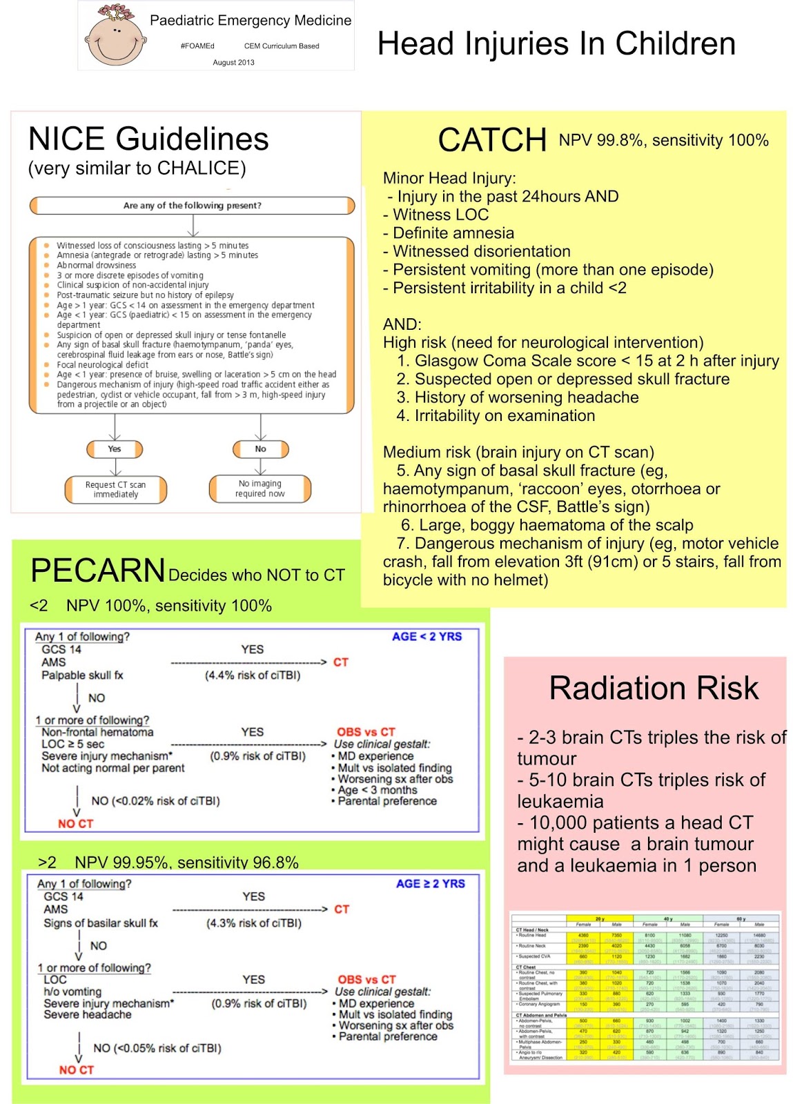
  • 2-3 brain CT triples the risk of brain tumours
  • 5-10 brain CTs triples the risk of leukaemia
  • Per 10,000 patients, a head CT might cause a brain tumour and leukaemia in one person. 
  • At 10 mSv Lifetime Attributable Risk of developing a cancer is 1:1000 
  • One additional cancer for every 4000 CT heads.  
So, as Clinicians we need to be able to balance the risk of a CT vs the risk of undiagnosed disease. Especially in children, the views of the parents often play a part in our decision making process, and maybe we should be gaining consent. If you'd like to be able to instantly show your patients their risk of cancer from a CT, there is a clear graphical display here, and there is a nice PV card here which unfortunately doesn't cover the paediatric population. X-ray risk has a more precise risk calculator, but again only estimates the risk in children.

And this is the risk if it all goes right! If it goes wrong, the doses could be a lot more.  And if there is a radiation emergency, what would you do?

Times are changing, and we're moving away from CT scans, and towards observation.  So think carefully before you do the CT scan.

All of this is nicely summarised by St Emelyns - there is often no right answer, and you have to balance the risks.


Saturday, 17 August 2013

Head Injuries

We know that the likelihood of children needing neurosurgical intervention after a head injury is slim

Head injuries in children can very easily fall into the grey area, where you're not sure whether the child needs a CT or not, and sometimes they fit the guidelines, but really don't need a CT... Page 9 of the NICE guidelines is very clear about when people need a CT, and there's a nice crib sheet from @_NMay here

The most debated point on the NICE guidelines seems to be "three or more discrete episodes of vomiting". The BMJ has investigated vomiting, and found it to be an unreliable sign. The SIGN guidelines report "repeated vomiting is a weaker predictor (RR 0.88) with reported OR ranging from 2.13 to 4.08 in three studies. In children, repeated vomiting had a PPv of 0.065". Yet they still suggest scanning these children. There's even a best bet saying vomiting isn't an accurate predictor although there is another best bet suggesting that persistent vomiting warrants a scan.

Children do vomit after an injury. If they don't get pain killers quickly enough, they vomit some more. They're prone to gastroenteritis and other bugs that make them vomit. And we don't like irradiating children because of the radiation risk. Trouble is, we're often happy to watch and wait, parents are often happy to watch and wait, but the admitting paediatric team (paediatricians, paediatric surgeons, neurosurgeons - who ever they come in under in your hospital) often want the reassurance of a normal CT scan before they'll admit. As Emergency Literature of Note notes, observation is often safer than a CT.

There are podcasts out there (and here and here) about head injuries.

There is another rule out there that is a lot more simple and doesn't include vomiting as a criteria - CATCH. Then there's CHALICE and the Kuppermann head CT rules, aka PECARN (summarised here, with a link to the original article and another podcast here).  These three rules are compared by the EMJ here, and online here. Importantly CHALICE and CATCH identify those who need a CT, and PECARN identifies those who do not:
PECARN
<2    NPV 100%, sensitivity 100%
>2    NPV 99.95%, sensitivity 96.8%

CHALICE
NPV 99.9%, sensitivity 98%

CATCH
NPV 99.8%, sensitivity 100% 

CATCH Rule (Canadian Assessment of Tomography for Children with Head Injury):
CT of the head is required only for children with minor head injury* and any one of the following findings:
High risk (need for neurological intervention)
1. Glasgow Coma Scale score < 15 at 2 h after injury
2. Suspected open or depressed skull fracture
3. History of worsening headache
4. Irritability on examination

Medium risk (brain injury on CT scan)
5. Any sign of basal skull fracture (eg, haemotympanum, ‘raccoon’ eyes, otorrhoea or rhinorrhoea of the CSF, Battle’s sign)
6. Large, boggy haematoma of the scalp
7. Dangerous mechanism of injury (eg, motor vehicle crash, fall from elevation 3ft(91 cm) or 5 stairs, fall from bicycle with no helmet)

*Minor head injury is defined as injury within the past 24 h associated with witnessed loss of consciousness, definite amnesia, witnessed disorientation, persistent vomiting (more than one episode) or persistent irritability (in a child <2 years of age) in a patient with a Glasgow Coma Scale score of 13/15.

CHALICE Rule (Children's Head Injury Algorithm for the Prediction of Important Clinical Events):
A CT scan is required if any of the following criteria are present.
  • History
    • Witnessed loss of consciousness of >5 min duration
    • History of amnesia (either antegrade or retrograde) of > 5 min duration
    • Abnormal drowsiness (defined as drowsiness in excess of that expected by the examining doctor)
    • > 3 vomits after head injury (a vomit is defined as a single discrete episode of vomiting)
    • Suspicion of non-accidental injury (NAI, defined as any suspicion of NAI by the examining doctor)
    • Seizure after head injury in a patient who has no history of epilepsy
  • Examination
    • Glasgow Coma Score (GCS) <14, or GCS <15 if < 1yearold
    • Suspicion of penetrating or depressed skull injury or tense fontanelle
    • Sign of a basal skull fracture (defined as evidence of blood or CSF from ear or nose, panda eyes, Battle’s sign, haemotympanum, facial crepitus or serious facial injury)
    • Positive focal neurology (defined as any focal neurology, including motor, sensory, coordination or reflex abnormality)
    • Presence of bruise, swelling or laceration >5cm if < 1 year old
  • Mechanism
    • High speed road traffic accident either as pedestrian, cyclist or occupant (defined as accident with speed > 40 m/h)
    • Fall of > 3 m in height
    • High speed injury from a projectile or an object
These are very similar to the NICE guidelines for CT head.

So are you going to scan your children or not? There isn't an easy answer, and your approach is going to be influenced by the injury mechanism, the clinical situation, your intuition, the trust policy and the guidelines above. At the moment, even in children, we seem to err on the side of caution, and if in doubt, scan. 

After you have scanned and discharged your children, or just discharged your children remember to give them some good quality head injury advice. The quality of advice leaflets is variable, and some of them make you look daft when they say return  straight away if there is any vomiting - and you've sent them home with three discrete guy episodes! I find it useful to encourage brain rest (no computers!) and headway has some excellent advice. For serious injuries, the Child Brain Injury Trust has some excellent parent  resources.

Parents often ask when their children can go back to sport. The brain is fragile and a second insult may be  more damaging. Consider the use of the "SCAT3" (Sport Concussion Assessment Tool).

There's a case for you to consider here, and a different case to contemplate.

Monday, 12 August 2013

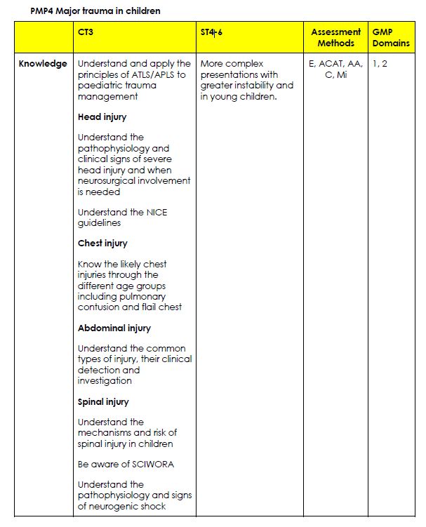
PMP4: Major Trauma in Children

This PMP has lots and lots of items in it. Having looked at this bit of the curriculum in detail, I'm skeptical as to whether attending APLS/EPLS really teaches you enough... What do you think?
Click here for all of the PMP4 / trauma posts.


Head Injuries. For a detailed head injury post click here, and for a detailed radiation risk post click here:


Chest Trauma in children. For more posts, click here.


For more posts on abdominal trauma, click here.

For more posts on burns, click here.
For posts on spinal trauma click here
For posts on pelvic trauma click here