Welcome

I struggled to find PEM resources for my CT3 year, despite the variety of excellent resources out there. I hope this website will help point you in the right direction. I'm not a PEM expert, but am following the guidance CEM have issued (in the form of a syllabus) to put together this page. This page is not endorsed by CEM, and any mistakes are mine.

Please comment with corrections, additions and further suggestions.

All the information here is collected from the internet, and it might be out of date or inaccurate, so please use your judgement and adhere to your hospital's protocols. If you do notice any errors or omissions please comment so we can put them right!

To navigate, decide whether you want to start with a PMP or a PAP. You can then select which PMP or PAP you want to look at. You will then be taken to the summary page for that PMP, with links expanded topic collections. If you know what topic you want to look at already, click on the link on the right hand side.
Showing posts with label PMP3. Show all posts

Thursday, 27 October 2016

Collapsed Neonates

Resuscitate
As per appropriate
Adrenaline 10mcg/kg

Prostin -  
    - 5 ng/kg/min if clinically well
    - 20 ng/kg/min if unstable or absent femoral pulses
    - 50-100 ng/kg/min if no response
Apnoea common: 1st hr of Rx, dose
Hypotension may occur with high dose

Prostin
- 5 ng/kg/min if clinically well
- 20 ng/kg/min if unstable or absent femoral pulses
- 50-100 ng/kg/min if no response
Apnoea common: 1st hr of Rx, dose

Hypotension may occur with high dose

Think about Causes
Infection
Group B strep, E Coli - PROM, maternal GBS, fever in labour
Herpes Simplex - GCS, coagulopathy, ALT, family cold sores
MRSA - Unresponsive 1st line antibiotics,+ contact

Cardiac
Coarctation aorta - Systolic arm/leg gradient > 20 mmHg
Hypoplastic Left heart - Poor pulses –may be pink= pulm. overcirculation
Transposition (TGA) - Preductal sats < post ductal sats
TAPVD (obstructed) - Shocked & cyanosed/CXR plethoric
SVT  - HR>220 despite fluid, f ixed HR, narrow QRS
Myocarditis - Cardiac failure, tachycardia, small QRS

Injury 
Intracranial bleed  - Focal neuro signs, fontanel le , retinal bleeds
Intrabdominal bleed - Unexplained anaemia, abdominal bruising

Cardiac

Metabolic
Vomiting, reduced GCS, hypoglycaemia
Stop the feeds. Give fluid and dextrose as highly likely to be fluid depleted

References
http://paediatricem.blogspot.co.uk/search/label/NLS
http://www.rcemlearning.co.uk/references/congenital-heart-disease/ 
http://www.rcemlearning.co.uk/modules/the-shocked-neonate/ 
http://www.rcemfoamed.co.uk/portfolio/metabolic-babies-in-the-ed-easy-as-1-2-3/ 

Monday, 12 August 2013

Sudden Infant Death (SIDs)

SIDS is defined as sudden and unexplained death in a child less than one year old, whose cause of death isn't immediately obvious prior to the investigation. SIDS is a smaller part of Sudden and Unexplained Infant Death - some of these are later found to have a cause. Over 300 babies die every year from SIDS. There is nothing we can do that guarantees protection from SIDS, but we can reduce the chances.

The Lullaby Trust offers specialist support for bereaved families and anyone affected by SIDs. They also have a leaflet with safe sleep advice for parents:
  •  Place your baby on their back to sleep. If they roll onto their front or side, roll them back again.
  • Keep your baby smoke free
  • Breastfeed your baby if you can. Even breastfeeding the baby some of the time can help reduce the risk of SIDs.
  • Place your baby to sleep in a separate cot or Moses basket in the same room as you for the first six months. 
  • Use a firm, flat waterproof mattress in good condition.
  • Using an orthodontic dummy after the age of four weeks MIGHT reduce the risk of sudden infant death.
Don't:
  • Never sleep on a sofa or armchair with your baby. 
  • Don't sleep in the same bed as your baby, especially  if you smoke, drink, take drugs or are extremely tired, or if your baby was born prematurely or was of low birth weight. 
  • Avoid letting your baby get too hot
  • Don't cover your baby's head or face whilst sleeping and use loose bedding. 

Management of SIDS depends on your hospital's protocol. Generally a senior Clinician (normally a Consultant Paediatrician) makes the decision to stop resuscitation. Specific protocols have to be followed and your hospital policies should be able to guide you on this. 

Cardiac Arrest Drugs

PMP 3 states we should:
"Understand the pharmacology, indications and contraindications, dose calculation and routes of administration of drugs used in resuscitation and the stabilisation of children in cardiac arrest."

Where better to start looking for resources on this kind of thing than in the anaesthetic literature?

Adrenaline
There is an anaesthetic FRCA tutorial here, which probably covers everything we need to know. It tells us similar things to LITFL. LITFL have a good summary here and here.
  • Naturally occurring catecholamine 
  • Hormone and a neurotransmitter
  • Broken down by monoamine oxidase (MAO) and catechol-O -methyltransferase (COMT).
  • Comes in 1:1000 strength (in vials) or 1:10000 (mini-jets)
  • Acts non selectively at all the adrenergic receptors (α1, α2, β1, β2, β 3)
    • At low doses the beta effects predominate - increase heart rate
    • At high doses the alpha effects predominate
  • Produces the flight or fight response (sympathomimetic)
  • Causes 
    • increased heart rate
    • increased contractility
    • vasoconstriction and vasodilation
    • bronchodilation
    • glycogenolysis
    • smooth muscle relaxation
  •  Side effects are normally because of overdose or inappropriate use. 
    • Dizziness
    • Palpitations
    • Tremour
    • Arrhythmias
    • Hypertension
    • Cerebral haemorrhage
    • Acute pulmonary oedema
    • Lactic acidosis
    • Hyperlactaemia - with long term use
    • Hyperglycaemia - with long term use
  • Caution
    • At all times: big effects on the cardiovascular symptoms
    • In patients with arrhythmias, hypertension or ischaemic heart disease
    • Care in patients taking Mono-Amine Oxidase Inhibitors
  • Uses
    • Cardiac Arrest: 1mg every 3-5 minutes, after 3rd shock. 10mcg/kg in children
      This is in the guidelines, but its use is still controversial
    • ROSC use 1: 100 000 to support BP if needed 
    • Anaphylaxis: Adult or > 12 500mcg IM or 50mcg IV titrated to effect if experienced in use.
      6- 12 years: 300mcg IM or 1mcg/kg IV titrated to effect
      <6: 150mcg IM or 1mcg/kg titrated to effect
    • Inotropic support: continuous infusion via central line with invasive BP monitoring
      • 4mg adrenaline diluted to 50ml in  saline or glucose
    • Severe croup: 
      •  0.5 ml/kg of a 1:1000 solution (maximum of 5 ml)
    • Topical or local vasoconstriction
      Remember:
      1ml of 1:1000 = 1 mg
      1ml of 1:10 000 = 0.1 mg = 100 mcg
      1ml of 1:100 000 = 0.01 mg = 10 mcg
Amiodarone
CV pharmacology has a good summary with some pretty pictures. LITFL has a good summary and a more pharmacology focused tutorial - they do state that efficacy of amiodarone in paediatric children hasn't been established.
  • Membrane stabilising anti-arrhythmic drug
  • Increases the duration of the action potential
  • Slows AV conduction
  • Mild negative inotropic effect
  • Causes peripheral vasodilation because of alpha-blocking effects
  • The solvent with it causes histamine release.
  • 25 - 60 days half life
  • Class I, II, III & IV actions
  • Generally considered a Class III arrhythmic drug
  • Dose:
    • 5mg/kg after third shock in shockable rhythms
    • Repeat the dose after the fifth shock
    • Central vein if possible - causes thrombophlebitis
  • Side effects
    • Pulmonary fibrosis
    • Thyroid problems
    • Can cause hypotension
    • Can cause Bradycardia and AV Block
    • Can cause liver enzyme elevations
    • May be arrhythmogenic
Atropine
There aren't so many summaries on atropine - probably because it's been taken out of the resus algorithm. There's something on CV pharmacology, LITFL summary and a page on the non resuscitation uses- Death Rattle.
  • Atropine is a muscarinic receptor antagonist and bind to muscarinic receptors. 
  • Stops ACh from binding
  • So stops the effects of vagal nerve activity on the heart
  • Administration of less than 500mcg in adults and less than 100mcg in children can produce a paradoxical bradycardia because of the central or peripheral parasympathomimatic effects of low dose.
  • Atropine rapidly crosses the placenta
  • Atropine crosses the blood-brain barrier
  • Side effects:
    • tachycardia
    • pupil dilation
    • dry mouth
    • urinary retention inhibition of sweating
    • blurred vision 
    • constipation
  • Contraindicated in patients with: 
    • glaucoma, 
    • pyloric stenosis and 
    • prostatic hypertrophy
  • Uses
    • Bradycardia - as per algorithm (adults and children 1 2 3)
      • May be caused by poisoning
      • Conduction disorder (pathway abnormality or Long QT)
      • Cardiomyopathy
    • Excessive secretions
  • Dose
    • 10mcg/kg
Magnesium
Magnesium is a panacea for everything at the moment. There is a summary article here, BNF entry here and a LIFFL summary. There are some good flashcards for Paramedics here. AAFP has got an interesting simply written article about magnesium.
  • Extracellular cation
  • Depressant effect on the release of catecholamines
  • Uses
    • Eclampsia
    • Torsades
    • Asthma
  • Contraindications
    • Heart block
    • Shock
    • Persistent hypertension
    • Hypocalcaemia
    • Myocardial damage
  • Cautions
    • Renal damage
    • Patients on digoxin
    • Use of CNS depressants or neuromuscular blocking agents
  • Side Effects
    • Hypotension
    • Sedation
    • Confusion
    • Bradycardia
    • Pruritis
    • Arrhythmias
    • Decreased deep tendon reflexes
    • Respiratory depression/paralysis
    • Drowsiness
    • Muscle weakness
    • Complete heart block
  • Dose
    • Torsade de Pointe
      • By intravenous injection over 10–15 minutes
      • Child 1 month–18 years 0.1–0.2 mmol/kg (25–50 mg/kg magnesium sulfate heptahydrate); max. 8 mmol (2 g magnesium sulfate heptahydrate); dose repeated once if necessary
    • Asthma
      • Children over 2 years with severe acute asthma may be helped by intravenous infusion of magnesium sulfate injection 40 mg/kg (max. 2 g) over 20 minutes
Calcium Carbonate
  • Used for: hyperkalaemia, hypocalcaemia, and over-dose of calcium-channel-blocking drugs
  • Dose for urgent calcium correction
    • By slow intravenous injection over 5–10 minutes
    • Neonate 0.11 mmol/kg (0.5 mL/kg of calcium gluconate 10%) as a single dose. [Some units use a dose of 0.46 mmol/kg (2 mL/kg calcium gluconate 10%) for hypocalcaemia in line with US practice]
    • Child 1 month–18 years 0.11 mmol/kg (0.5 mL/kg calcium gluconate 10%), max 4.5 mmol (20 mL calcium gluconate 10%)
Sodium Bicarbonate  
From the PALS algorithm:
"The best treatment for acidaemia in cardiac arrest is a combination of effective chest compression and ventilation (good quality CPR). Administration of sodium bicarbonate generates carbon dioxide, which diffuses rapidly into the cells, exacerbating intracellular acidosis if it is not rapidly cleared via the lungs.
It also has the following detrimental effects:
  • It produces a negative inotropic effect on an ischaemic myocardium.
  • It presents a large, osmotically active, sodium load to an already compromised circulation and brain.
  • It produces a shift to the left in the oxygen dissociation curve further inhibiting release of oxygen to the tissues.
The routine use of sodium bicarbonate in cardiac arrest is not recommended. It may be considered in prolonged arrest, and it has a specific role in hyperkalaemia and the arrhythmias associated with tricyclic antidepressant overdose. "
  • Dose

    • By slow intravenous injection of a strong solution (up to 8.4%), or by continuous intravenous infusion of a weaker solution (usually 1.26%)
    • An amount appropriate to the body base deficit


Friday, 9 August 2013

Electrocution in Children

Electrocution is surprisingly common, although rates of lightening strike are decreasing. Given that they should be well aware of the dangers of electricity, I was surprised that electricians and people who work with electricity are most likely to be affected. Electrocutions could affect people in hospital as well as out of hospital, and as the old adage goes, prevention is better than cure. It's not just high voltage electrocutions that cause problems, as a 33 year old with a domestic electrocution found.

If you look hard enough, you can find plenty of information online about electrocution. The AHA has a very thorough article online and EnlightenMe and LITFL have cases on the injuries associated with electrocution.
I know it's not FOAM, but the eLfH e-learning site has a fantastic e-learning module about electrical burns. It tells you a lot about those things that you always wondered about, but weren't quite sure about, and I'd really recommend completing it if you can.

So, lets get started.

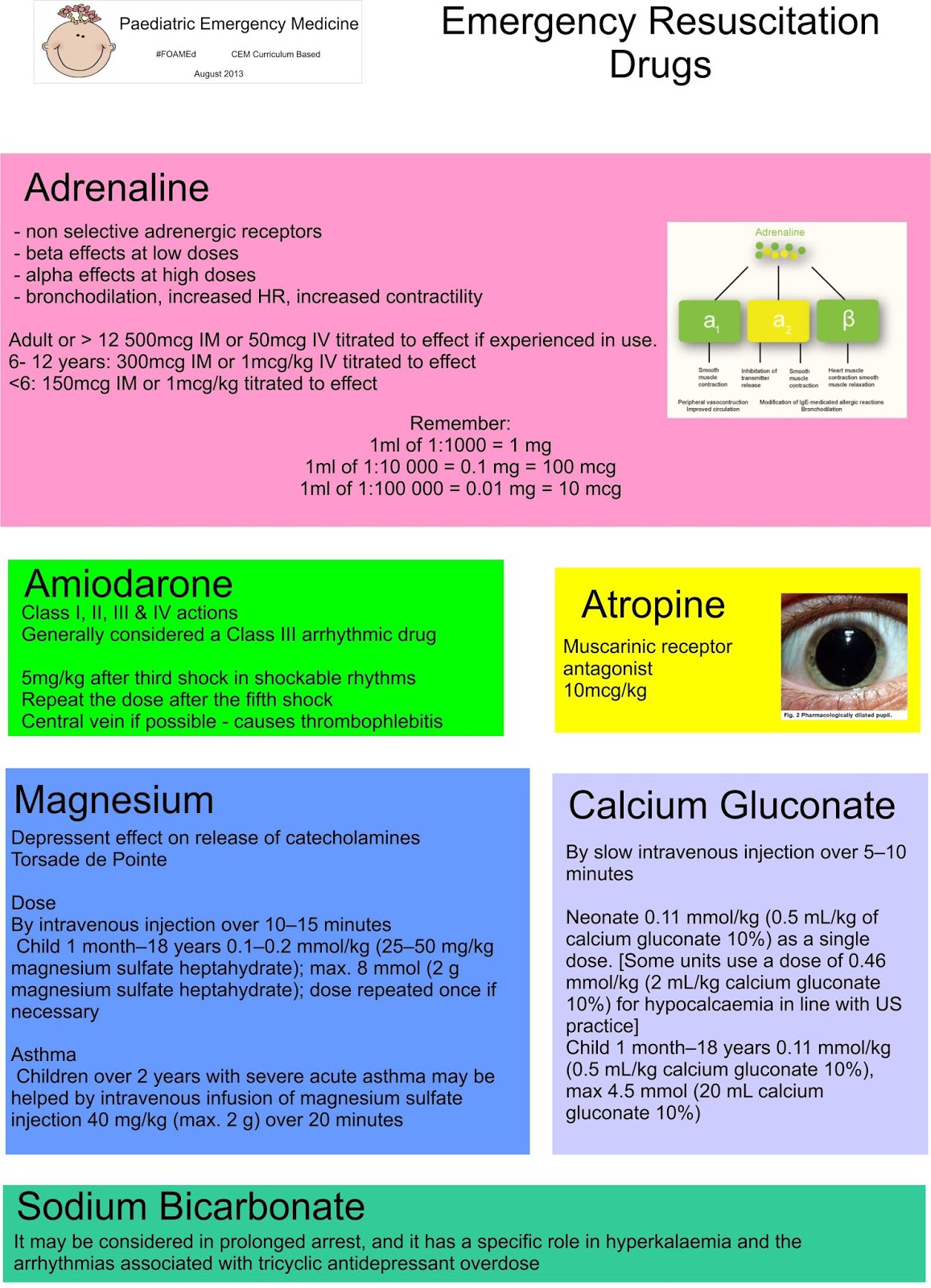
Electrocution: death as a result of exposure to electric current

Electrical injury: Tissue damage as a result of exposure to electrical current
Electrical shock: The violent response to electric current exposure often characterised by involuntary muscle contraction.

1000 people per year are estimated to die from exposure to electricity each year. In 2005, there were 11 work related electrocutions and 119 serious injuries in the workplace. Electrical injuries follow a bimodal age distribution pattern - children younger than six, and in early adulthood.


The ability of an electrical current to cause morbidity and mortality is dependent on six different factors:
- Magnitude of the current
- Voltage of the electrical source
- Resistance of tissue types involved - (bone has the most resistance)
- Duration of exposure
- Type of current
- Current pathway

There are two main types of electricity that might cause us injury - low tension and high tension.
 
Low Tension
  • <300 Volts 
  • at 50hz  
  • Alternating current  (AC)
  • Domestic (110V in the US and Canada; 220V in Europe, Australia and Asia).
  • Can cause VF
  • Theoretical risk of late VF if shock travels through the thorax
    May cause tetanic skeletal muscle contractions - stopping people from letting go from the electricity source. Because AC current is repetitive it also increases the likelihood of current flow through the heart during the relative refractory period of the cardiac cycle which can precipitate ventricular fibrillation.
  • Intraoral burns are common in young children (toddlers who explore electrical cables by placing them in their mouth). Conduction may be aided by electrolyte rich saliva. Delayed haemorrhage is a recognised feature. 
    Prevention is always important, and ROSPA have lots of advice (and lots of leaflets) about how to make your home safer.

High Tension
  • >1000 Volts
  • Lightening strikes and overhead power cables.
  • Direct current
  • May cause asystole, complete heart block and QT prolongation. 
  • Keraunoparalysis - transient limb weakness, rarely lasting more than a few hours. Upper limbs > lower limbs. 
  • Lightening strikes - mortality of 30%
    • extensive autonomic stimulation - hypertension, tachycardia and nonspecific electrocardiographic changes
    • brain haemorrhages, oedema and nerve injury.
  • Lichtenberg’s flowers 
    • Transient, fernlike erythematous floral pattern that develops on the skin
    • Fades in 24 - 36 hours 
This type of electricity simultaneously depolarises the entire myocardium. This normally stops the heart completely, but sometimes the heart sorts itself out again and starts working normally again (it has intrinsic cardiac automaticity). This type of electricity also causes thoracic muscle spasm making it very difficult to breathe. Secondary hypoxic cardiac arreast is likely! Victims are most likely to die if they experience immediate respiratory or cardiac arrest and no treatment is provided - if treatment is provided at scene, results are often good. 
DC exposure is single, and often throws the victim away. A direct strike is uncommon, and often splashes sideways from an object or a victim may be holding on to a struck object.

Treatment
  • Early basic life support and defibrillation if needed
  • Airway:
    • Secure the airway early (airway burns likely)
  • C-Spine Control:
    • Especially if DC current - may have been ejected
  • Breathing:
    • AC current may paralyse thoracic muscles
  • Circulation:
    • Hypovolaemia from concurrent injuries likely
    • ECG - assess for presence of sinus rhythm, and remember to check the QT interval
    • Cannulate in a limb not involved in the current pathway 
      • There may be damage to vascular structures that will stop systemic circulation
  • Disability: 
    • Look for entry and exit burns
    • Cool burns as appropriate 
    • Remove smouldering clothes, shoes and belts. 
  • Exposure: 
    • Look for compartment syndrome (if you don't look you won't see!)
    • Bloods for:
      • FBC, U&E (hyperkalaemia secondary to rhabdomyolysis likely)
      • Phosphate
      • Calcium
      • CK
      • Group and Save
      • Role of troponins is unclear 
    •  Check hearing
      • Risk of ruptured ear drums from lightening (Oxford Handbook of Emergency Medicine)
    • Urinalysis - check for blood
  • Observe
    • Six hours of being asymptomatic
  • Send home
    • Asymptomatic patients with 
      • domestic and minor low voltage burns
      • normal ECG
      • no history suggestive of arrhythmia
      • no myoglobinuria
  • Admit
    • Children who bite electrical flexes
      • risk of delayed bleeding
    •  All patient with high voltage conduction injuries, or abnormalities found.
  • Follow Up 
    • Delayed cataract formation 
    • Neuropsychiatric problems
    • Compartment syndrome advice
The management of burns is important - we'll cover this later!

Tuesday, 6 August 2013

Venous Access in Children

Intravenous Cannulation
 Most of us are skilled at gaining IV access in adults, but get a bit more concerned when we need to bleed or cannulate a small child. Here's a few tips I've picked up on the way...

Preparation and Distraction
  • Get all your stuff ready and open before you start. This means the rustling isn't off putting.
  • If the child is big enough, sit them on a parent's knee, and position the hand behind them, so they can't see what is going on. 
  • Distraction is really important - parents are excellent at this. They might use an iphone, Where's Wally, or singing of Nursery Rhymes.
  • Go in to the room feeling positive and as though you WILL succeed - children pick up on the vibes.
  • Ametop or Emla cream is very helpful if you have time.
  • Babies still feel pain - sucrose can be helpful. 
  • If you don't have time for Ametop to work, remember cold spray can be helpful. 
  • Analgesic gas can also be very useful - using it also distracts the child!
Technique
There are plenty of excellent resources demonstrating a good technique.
From personal practice, I think the most important steps are distraction (otherwise you have a wriggling child), confidence, and holding the skin really really taut before attempting the cannulation.
What are your tips?

Intraosseous Cannulation
IO is a new idea in adult trauma, but has been used in children for a while. There's a great article on IO in trauma here. There are many methods of gaining IO access in children and despite them all seeming as good as each other in adults,  EZ-IO seems better and quicker in children, with low complications... Whether your department uses a Cook needle or a gun type device like the EZ IO make sure you know how to use it, and which needle to use. Most devices use a pink needle for children.

Although it doesn't alter flow rates, the site you chose for IO access is really important in children, as you don't want to damage their growth plates. These guidelines have clear location pictures reminding us of where the growth plates are. EZ - IO have a clear powerpoint with guidelines too. 
  1. Tibia
    Find the flat anteromedial surface of the tibia one- to two-finger breadths below the level of the tibial tuberosity is the preferred site.
    Position the leg with the knee slightly bent and semi-externally rotated. Place a sandbag or towel roll under the leg for support.
  2. Ankle
    One- to two-finger breadths proximal to the medial malleolus, sufficiently posterior to avoid the saphenous vein.
    Externally rotate the leg.
    Use only as last choice in young children
  3. Humerus
    1 cm above the surgical neck (where the bone juts out)
  • Clean the area
  • Insert the IO
  • After you have inserted the IO, you need to unscrew the needle from the stylet, and dispose of the needle. If you forget to unscrew the needle your IO won't work!
  • Secure the IO 
  • Consider  flushing with local anaesthetic.
  • Aspirate bone marrow for analysis (and remember to let the lab know).
  • Attach extension tubing to the IO. This means you can easily access the IO without repeatedly moving the IO.
Keep practicing when ever you can on the non-poorlies, and then when you need to gain IV or IO access in a poorly patient, you'll have no problems.

Hypothermia

We're all used to deliberately inducing hypothermia in patients who have had an arrest, or similar - but not so used to treating hypothermia. For a good environment based start to your hypothermia training, there are plenty of Wilderness Medicine and Polar Medicine courses about.

EnlightenMe has got a lot of resources on hypothermia. Most of them are about adults, but that is better than nothing! e-LfH has a module on hypothermia too. We have some learning about ECG changes, a CEMPedia article, CEMPedia on cardiac arrest in special circumstances. BMJLearning has two modules - hypothermia for Paramedics and accidental hypothermia which has an excellent list of things that predispose people to hypothermia. LITFL also talks about ECG changes, as does Circulation- the height of the Osbourn wave is proportional to the degree of hypothermia.

There is a podcast here, a LITFL summary, and an abbreviation heavy summary here on crashing patient. There is an excellent summary here on Academic Life in Emergency Medicine. There are also some well thought of videos here.

For those who are wondering whether it's all worth it or not, there is a brilliant video about a hypothermia survivor.

Statistics
In Great Britain, hypothermia cases are estimated at 6-8 patients per 1000 patients Most cases of hypothermia occur in an urban setting and are related to environmental exposure attributed to alcoholism, illicit drug use, or mental illness, often exacerbated by concurrent homelessness.

Primary hypothermia usually affects young males and infants. Secondary hypothermia usually affects patients who are elderly, homeless, mentally ill, victims of trauma, or have multiple co-morbidity.

Older adults are at risk of hypothermia as a consequence of:
  • A reduced ability to recognise and respond to lower ambient temperature, compared with younger adults. 
  • An impaired peripheral vasoconstrictor response to cold.
  • A lower basal metabolic rate compared with younger adults. 
  • Falls, which are common in older people can result in prolonged periods lying on the floor, allowing heat loss by conduction.
Signs and Symptoms
There are plenty of signs and symptoms of hypothermia - some of which are subtle, others aren't.
Shivering
Impaired judgement
Confusion
Tachycardia
Tachypnea

Bradycardia
Respiratory depression
Hyperglycaemia
Dysarthria
Ataxia

Stupor
Lethargy
Osbourne Waves on the ECG - ( Osborn or J waves on the ECG. The upward deflection of the terminal S wave)
Loss of reflexes
Hypotension
Asystole

Treatment
  • ABCD approach
  • Handle carefully
    • In general, the hypothermic patient is dehydrated and fluid depleted consider a fluid challenge of warmed 0.9% saline or dextrose-saline as they may also be hypoglycaemic. Hartmann’s is best avoided as the hypothermic liver can't metabolise lactate. 
  • Passive Rewarming
  • Active Rewarming
    • Core Rewarming - IV fluids and warm, humidified oxygen
    • Extracorporeal Blood Rewarming
In cardiac arrest
 - Total of three shocks (if indicated) until core temperature greater than 30degrees.
 - No drugs under 30degrees, then given with twice the time interval until normothermia
 - Chest compressions may be harder work.

Investigations
Urea & electrolytes
Full blood count
Clotting screen,
Arterial blood gas
Creatinine kinase level
Calcium
Magnesium
Amylase

Drowning

Drowning and near drowning is a fascinating area. As a dinghy sailor, I regularly teach about drowning on our first aid courses, but as we sail on top of the water and not in the water, I've still never seen it! If you get a chance to watch the Cold Water Casualty video, it is well worth a watch. The Royal Yachting Association has a good summary about cold shock, hypothermia and drowning. The RNLI provides some UK statistics. Drowning is specifically mentioned in the cardiac arrest competencies, and in HAP11 Environmental emergencies (an adult competency).

WHO provides us a statistical summary about drowning, and Update in Anaesthesia provides a very thorough overview as does BMJ best practice, LITFL, EP monthly and the AHA.

There is an e-learning module on drowning here and here. Unless you are a Paramedic you're unlikely to have access to the former, and there are so many alternative resources out there I wouldn't subscribe. Enlighten me has an excellent CEMPedia article on drowning  and an adult case.


Statistics
Drowning is the process of experiencing respiratory impairment from submersion/immersion in liquid. In 2000, there were 409,272 deaths from unintentional drowning worldwide, decreasing slightly in 2004 to an estimated 388 000. 150 people drown in the UK coast every year - and 80% of these are male. Drowning is the second highest cause of death from injury, after road traffic injuries.

Young children are at risk as they are unaware of the dangers and less able to escape from water once submerged.

Things Drowning Causes
There are four main problems drowning causes:
  • Hypothermia and Cold Water Shock
    Not everyone drowns in warm water! Hypothermia causes its own special problems. The most relevant problem is that blood gets diverted in to the core of the body - causing chilly blood outside, and warm blood inside. When you start to mix the warm and chilly blood, the body gets a bit confused - and arrhythmias can occur.
    Cold shock can happen at any water temperature below fifteen degrees - the average UK water temperature is twelve degrees.
  • Associated injuries
    Remember to think about what people injured when they drowned themselves - neck and c-spine injuries are common.
    Awareness of injuries will be low because of natural adrenaline, and vasoconstriction in the extremities.
  • Water aspiration
    As you hit the water, before the diving reflex is triggered, you get a gasp reflex (as you do when you first get in a really cold swimming pool!). If you enter turbulant water, as waves crash overhead you are even more likely to inhale some water. Aspiration of even small amounts of water significantly impairs gas exchange. The water dilutes and inactivates surfactant, so alveoli are prone to collapse. The water can also cause direct lung injury.
  • Post rescue Collapse
    Loss of hydrostatic pressure of water on the body results in loss of peripheral resistance and venous pooling. Patients should be extricated from the water in a horizontal position.
  • Post Drowning Infection
    Some people contract infections after drowning. Swamp water might cause fungal infection (aspergillosis)- so think about it in your poorly patients. Stagnant water with rat urine may cause Weil's disease (Leptospirosis). There are many case reports (1 2 3) about pneumonia after drowning - but there is still no evidence for prophylactic antibiotics.
Investigations
- ABG if significant history of submersion as saturations may not be reliable.
- CXR
- ECG - look closely for a prolonged QTc.
- Core temperature measurement
- Electrolytes and BM
- Blood culture in patients with significant aspiration

Treatment
  • ABCD approach
  • For children, no modifications to the resuscitation sequence are recommended.
  • If intubation needed, ventilate using an ARDS protocol. 
  • Observe for  at least six hours.
  • After discharge, written discharge advice is well received by families. 
  • There is no evidence to support giving prophylactic antibiotics on discharge.


Cardiac Arrest in Children - aetiology

There are two types of cardiac arrest in adults, and children.

Primary Cardiorespiratory Arrest: due to an underlying cardiac arrhythmia (eg VF or VT) is more frequent in adults. The onset is often acute and unpredictable. Immediate defibrillation is needed.

Secondary Cardiorespiratory Arrest is more common in children and is because the body can't deal with the underlying injury or illness.
The pre-terminal rhythm in children is often bradycardia which leads to asystole or PEA - non shockable rhythms.

The outcome from cardiorespiratory arrest in children is poor, especially if there is a prolonged duration. Compared to adults, children are physiologically different but like adults, early recognition of the seriously ill child can prevent sudden death.

Some conditions are more likely to affect children than adults, and more likely to cause their arrest:
Croup: remember a narrower tracheal tube than would normally be expected may be required.
Epiglottitis: you must keep the child calm until the airway is secure.
Bronchiolitis
Coma
Seizures
Anaphylaxis: give adrenaline as quickly as possible

Drowning, electrocution and hypothermia can also affect children, and cause cardio-respiratory arrest. In an arrest situation, the standard CPR algorithms should be followed.

Survival rates remain poor, with survival rates of 0 - 38% quoted in the literature.

Monday, 5 August 2013

Paediatric Resuscitation

Most people learn by doing, whether their "doing" is simulation, or real life. Given the thankful infrequency of paediatric arrests, we must rely on simulation. I would strongly recommend you practice paediatric resuscitation simulations, making them as realistic as possible, to supplement your theoretical knowledge. I've heard that one children's hospital runs a mock paediatric arrest weekly, and it is very helpful.

Course wise, either APLS or EPLS is accepted. Most people I've spoken to prefer the APLS course as they syllabus is broader. I've not done both of them so I can't comment, but I enjoyed my course.

Guidelines
American Heart BLS guidelines
American Heart ALS guidelines
American Heart ALS and BLS
American Heart Summary
Anaethetists Update
Resus Council Guidelines

Issues
This blog has a look at what goes wrong in a paediatric arrest. With a link to a formal study, they conclude that "issues regarding equipment familiarity/use/misuse, failure to check BSL, and drug errors. Calculations of drug doses were difficult under stress. Failure rates in some of these domains exceeded 50%.

Drug dosing is difficult. Multiple smart phone apps exist - I like PalmPedi, despite the American-ness (if you buy PalmPedi consider getting PalmEM - reviewed on LITFL instead, as PalmED encorporates PalmPedi). This Australian website is nice and clear. We'll blog about drugs later.

Word of mouth suggests that "WET FAG" is no longer used as an acronym, and "WET FLAG" is more common instead. I do try and memorise all of these doses, but do like to be able to swiftly check them. I do not trust my phone to have signal or battery, and other Clinicians are often busy - so I rely on paper versions instead.

Parents - in or out?  In my experience they want to be in, and it is useful to have someone explaining things to them. They don't alter efficacy of the resuscitation effort.

Paeds arrest in trauma? Survival rates aren't good.

And debriefing afterwards is good, even if the resus is successful. Remember to carefully document - especially as you may have caused rib fractures, which may be later attributed to non accidental injury.

The Algorithm
Apart from doses, the algorithm is pretty much the same as for adults with a few key differences:
 - Start with 5 initial rescue breaths
 - CPR ratio 15:2

There are plenty of similarities:
 - No atropine unless bradycardic
 - Sodium bicarbonate only recommended if prolonged arrest, or associated with hyperkalaemia or TCA overdose
 - Continuous compressions encouraged.
 - After ROSC, titrate oxygen to saturations


Newborn Life Support

Having to perform NLS is rare, but is an easy exam scenario. The NLS course is a very useful one day course to attend. If you can't get on an NLS course, I'd recommend reading the NLS course manual - it has many useful pictures and tips. It is worth remembering that the N in NLS stands for "Newborn" and NOT "Neonatal". This means that if you have a poorly baby who is even six hours old, they are not a new born, so APLS guidelines should be used, not NLS.

If you look hard enough, there are quite a few neonatal life support resources.
The resus council protocol is here: Newborn Life Support.
The American Heart Association guidelines are here.

Life in the fast lane have a very useful (no pictures, so quick to load) summary here with the SMACC conference video on newborn resuscitation. AAFP also has guidelines here.  PedEMMorsels has a very matter of fact summary here. The anaesthetists have had their own update here. WikiEM also has a very thorough summary. Academic life in EM has some good videos, supplemented with pictures of stick men!

Make sure you know your hospital's protocols. Many hospitals have a seperate Neonatal resuscitation team, as well as the Paediatric resus team.
If you have a resuscitaire in your department, check that you know how to use it. If you don't have a resuscitaire, where is your nearest?


And if you were wondering - yes, you can use an LMA in new born resus - sanscrit and resus.me have the answers.

PMP3: Cardiopulmonary Resuscitation

Paediatric Arrests don't happen often - but when they do we should be ready. The curriculum and summaries are on this page.
To have a look at the detailed posts and links for each topic, please go here.


The ALiEM summary on hypothermia is so good there's no point reinventing the wheel.For more hypothermia blog posts, please click here.

For more NLS blog posts, please click here.


For more PLS blog posts, please click here.


For more electrocution blog posts, please click here


For more blog posts on arrest drugs, please click here.


There are also posts on:
Venous Access 
SIDS
Drowning