Welcome

I struggled to find PEM resources for my CT3 year, despite the variety of excellent resources out there. I hope this website will help point you in the right direction. I'm not a PEM expert, but am following the guidance CEM have issued (in the form of a syllabus) to put together this page. This page is not endorsed by CEM, and any mistakes are mine.

Please comment with corrections, additions and further suggestions.

All the information here is collected from the internet, and it might be out of date or inaccurate, so please use your judgement and adhere to your hospital's protocols. If you do notice any errors or omissions please comment so we can put them right!

To navigate, decide whether you want to start with a PMP or a PAP. You can then select which PMP or PAP you want to look at. You will then be taken to the summary page for that PMP, with links expanded topic collections. If you know what topic you want to look at already, click on the link on the right hand side.
Showing posts with label ct. Show all posts

Saturday, 7 September 2013

Abdominal Trauma


The abdomen is the third most injured body part in children, after the head and the chest.

There is a medscape e-learning module here, and a good pdf overview here, here, and another overview here with a summary on page 13. There is some discussion around a case here and some good CT images here. There is an excellent summary about abdominal trauma in children here, which introduces us to some guidelines for careful imaging in children.The anaesthetists at Lewisham wrote a very thorough overview here. There's a good summary of common injuries and their management here.

There is a good CT scan with some SAQs from the PMJ here.

Anatomical differences in children:
- The abdomen is square and becomes more rectangular as the child matures.
- Muscles are thinner, so there is less protection for underlying structures.
- Ribs are more flexible so they are less effective at energy dissipation, so less effective at protecting the upper abdominal structures.
- Solid organs are comparatively larger so are at more risk for injury.
- Attachments are more elastic.
- The intestine is not fully attached within the peritoneal cavity so is more vulnerable to injury due to sudden deceleration and/or abdominal compression.
- The bladder is more exposed.
- Children's spines are exposed to chance fractures, especially if they are restrained with a lap belt only.
- Abdominal distension from aerophagia is common
- Hypothermia is more likely

Clinical Features
The Seatbelt Sign
The seat belt sign is a good indicator of serious injury. In one review, 78% of patients with a seat belt contusion had intra-abdominal injuries, although all children with injuries also had abdominal pain - not just a seat belt sign.
Lap belts are designed to be worn at or below the anterior superior iliac spine level. In smaller children the belts are in the wrong place, and children can move out of them very easily. The introduction of booster seats has helped to improve this. You are up to 3 times more likely to be injured if you are not properly restrained.

Haematuria
The most common indication for abdominal imaging after trauma in children is reported to be haematuria. Non–urinary tract injury is observed more frequently than urinary tract injury in children with haematuria and asymptomatic hematuria is a low-risk indicator for abdominal injury.

80% of injuries are from blunt mechanism.

Holmes 2012 - decision Rules
Holmes looked at children with blunt torso trauma, and suggested some decision rules for when we should be CTing these patients. The paper has been reviewed on PEMLit. He has listed seven factors that make the injury low risk:
Most patients I've seen with trauma do complain of some abdominal pain -so maybe one or two CTs might be saved! This has a 99.9% negative predictive value.

If we do CT it's pretty good at picking up pathology.  If the patient is stable, ultrasound and serial observation is probably sufficient. There's a suggestion of an algorithm here. In a verbal patient, normal obs, normal examination and no abdominal pain is a pretty good indicator of no abdominal injury.

Wednesday, 21 August 2013

Chest Trauma in Children

"Know the likely injuries through different age groups including pulmonary contusion and flail chest"

Isolated significant chest trauma is rare in paediatrics, but is still the second highest cause of trauma related death in paediatrics (although whether the death was caused by the chest or by other injuries is difficult to tell). There is an excellent handbook from the Royal Children's Hospital in Melbourne which probably  tells you everything you need to know. There is a 2008 review in "Trauma" and a really nice overview presentation here.

Most chest injuries are caused by blunt trauma and  RTCs, and are normally associated with injuries elsewhere. The anatomy is similar to adults (excellent podcast here), but children are different - especially if they are younger:
  • Smaller
    • Traumatic forces distributed over a smaller mass
    • Smaller thoracic volume
    • Compact organs
  • Increased chest wall compliance
    • Ribs unlikely to break
    • Internal injuries likely with minimal external signs
  • Compensatory mechanisms may mask hypovolaemia
  • Mobile mediastinum
    • Airway injuries rare
    • Mediastinal shift may cause problems
  • Aerophagia is a common response in children
    • Causes reflex ileus
    • Leads to gastric dilation 

As in adults, there are six life threatening chest injuries to remember, remembered by the mnemonic "ATOM FC". These should be identified on the primary survey:
"ATOM FC"
 - Airway Obstruction
 - Tension Pneumothorax
 - Open pneumothorax
 - Massive haemothorax
 - Flail chest
 - Cardiac Tamponade

The Trauma journal review also reports there are six "hidden" but life threatening injuries:
-Cardiac contusions
-Aortic disruption
-Tracheobronchial disruption
-Oesophageal disruption
-Diaphragmatic tear
-Pulmonary contusion

We're hopefully pretty good at identifying the life threatening injuries. Often we suspect the "hidden" injuries. But how do we make the final diagnosis, especially as we know that we don't want to do unnecessary CTs on children.

This short presentation introduces some clinical decision rules to help you decide who needs chest imaging. Using a paper from the Annals of Emergency Medicine (which I can't access as it doesn't support Athens or Institutional Access) they suggest we only do a CXR in trauma if:
  • Low BP/Increased RR
  • Abnormal chest exam
  • GCS<15
  • Femur fracture
After trauma, most children I've seen have got an increased respiratory rate, so making sure we do a CXR for everyone doesn't seem unreasonable.  What about CT?

Pediatric Radiography have looked at how CT changes our management of paediatric patients. Again, I can only access the abstract:
235 children had a CXR and a CT done. In 1/3 CXR abnormal, and in 2/3 CT was abnormal.
Out of all the children, <5% got any chest procedure

<10% of kids with pneumothorax on CT got a chest tube
91% of CT findings were within 1cm of dome of liver --> visible on CT abdomen.

So this study would suggest that routinely doing a CT chest on all children may be unnecessary. Given that most of our treatment for chest injury is analgesia, and supporting ventilation, CT rarely changes management. Not all pneumothoraces are treated with a chest drain.
I'm not sure I'd be ready to chance hospital policy yet, but it is certainly something to think about! 

Lung Contusion
This is the most common chest injury in children, and can occur without chest wall injury.

Diagnosis:
-  Hypoxia
-  Consolidation on CXR (in 90% on initial CXR)
        - May be irregular and not conform to lobes
- CT scans pick up more scans
       - 38% of dogs experiencing blunt trauma had consolidation on CXR, compared to 100% on CTs


Treatment:
- Oxygen
- Analgesia
- Pulmonary toilet
- Ventilatory Support

Flail Chest
Flail chest is very rare, but can happen. The paraxodical chest movement caused by two or more rib fractures contributes towards respiratory distress. These often occur with pulmonary contusions. There is a very good explanation with some excellent pictures here.

Treatment
- Place the patient injury side downwards
- Intensive respiratory support
- Analgesia

Sunday, 18 August 2013

Radiography Risk

There's lots of data about the dangers of CT scans, and we certainly do a lot of scans in the Emergency Department. Although not specifically mentioned in the CEM syllabus, I think it is an unspoken part of every test we request, especially head CTs. I don't think we quite "order numerous CT scans each shift, and dole out radiation exposure dwarfing the atomic bomb dropped on Hiroshima" but we are getting closer than we used to! Just as the sports medics are reducing the number of scans they do, so do we need to.

If you're like me and don't know a lot about radiation, PEMMorsels has a good introduction, as does the Short Coat:
  • 1 Sv= 100 rem.  We use milliSieverts in most medical imaging.
  • Neoplasms have typically been associated with radiation in excess of 50 mSv
  • Background radiation = 3.5 mSv/year 
  • Background Cancer Incidence = 420:1000 (42%)
  • Medical radiation is between 0-100 mSv
 Although x-rays have a radiation risk, we worry more about the risk of CTs - as summarised on LITFL:
  • Routine abdominal CT scan estimated exposure quoted as 8-10 mSv
  • Actual calculated radiation: Average 11-20 mSv (Range 4-45 mSv)
  • Multiphase abdomen and pelvis CT scanning: median 31 mSv (Range 6-90 mSv)
  • Chest CT for suspected pulmonary embolus: median 10 mSv (Range 2-30 mSv)
  • Routine head CT median:  2-3 mSv (Range 0.3-6 mSv)
With these radiation doses, we might easily be causing pathology:
  • 2-3 brain CT triples the risk of brain tumours
  • 5-10 brain CTs triples the risk of leukaemia
  • Per 10,000 patients, a head CT might cause a brain tumour and leukaemia in one person. 
  • At 10 mSv Lifetime Attributable Risk of developing a cancer is 1:1000 
  • One additional cancer for every 4000 CT heads.  
So, as Clinicians we need to be able to balance the risk of a CT vs the risk of undiagnosed disease. Especially in children, the views of the parents often play a part in our decision making process, and maybe we should be gaining consent. If you'd like to be able to instantly show your patients their risk of cancer from a CT, there is a clear graphical display here, and there is a nice PV card here which unfortunately doesn't cover the paediatric population. X-ray risk has a more precise risk calculator, but again only estimates the risk in children.

And this is the risk if it all goes right! If it goes wrong, the doses could be a lot more.  And if there is a radiation emergency, what would you do?

Times are changing, and we're moving away from CT scans, and towards observation.  So think carefully before you do the CT scan.

All of this is nicely summarised by St Emelyns - there is often no right answer, and you have to balance the risks.


Saturday, 17 August 2013

Head Injuries

We know that the likelihood of children needing neurosurgical intervention after a head injury is slim

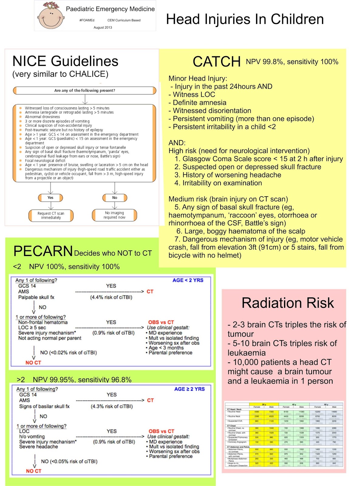
Head injuries in children can very easily fall into the grey area, where you're not sure whether the child needs a CT or not, and sometimes they fit the guidelines, but really don't need a CT... Page 9 of the NICE guidelines is very clear about when people need a CT, and there's a nice crib sheet from @_NMay here

The most debated point on the NICE guidelines seems to be "three or more discrete episodes of vomiting". The BMJ has investigated vomiting, and found it to be an unreliable sign. The SIGN guidelines report "repeated vomiting is a weaker predictor (RR 0.88) with reported OR ranging from 2.13 to 4.08 in three studies. In children, repeated vomiting had a PPv of 0.065". Yet they still suggest scanning these children. There's even a best bet saying vomiting isn't an accurate predictor although there is another best bet suggesting that persistent vomiting warrants a scan.

Children do vomit after an injury. If they don't get pain killers quickly enough, they vomit some more. They're prone to gastroenteritis and other bugs that make them vomit. And we don't like irradiating children because of the radiation risk. Trouble is, we're often happy to watch and wait, parents are often happy to watch and wait, but the admitting paediatric team (paediatricians, paediatric surgeons, neurosurgeons - who ever they come in under in your hospital) often want the reassurance of a normal CT scan before they'll admit. As Emergency Literature of Note notes, observation is often safer than a CT.

There are podcasts out there (and here and here) about head injuries.

There is another rule out there that is a lot more simple and doesn't include vomiting as a criteria - CATCH. Then there's CHALICE and the Kuppermann head CT rules, aka PECARN (summarised here, with a link to the original article and another podcast here).  These three rules are compared by the EMJ here, and online here. Importantly CHALICE and CATCH identify those who need a CT, and PECARN identifies those who do not:
PECARN
<2    NPV 100%, sensitivity 100%
>2    NPV 99.95%, sensitivity 96.8%

CHALICE
NPV 99.9%, sensitivity 98%

CATCH
NPV 99.8%, sensitivity 100% 

CATCH Rule (Canadian Assessment of Tomography for Children with Head Injury):
CT of the head is required only for children with minor head injury* and any one of the following findings:
High risk (need for neurological intervention)
1. Glasgow Coma Scale score < 15 at 2 h after injury
2. Suspected open or depressed skull fracture
3. History of worsening headache
4. Irritability on examination

Medium risk (brain injury on CT scan)
5. Any sign of basal skull fracture (eg, haemotympanum, ‘raccoon’ eyes, otorrhoea or rhinorrhoea of the CSF, Battle’s sign)
6. Large, boggy haematoma of the scalp
7. Dangerous mechanism of injury (eg, motor vehicle crash, fall from elevation 3ft(91 cm) or 5 stairs, fall from bicycle with no helmet)

*Minor head injury is defined as injury within the past 24 h associated with witnessed loss of consciousness, definite amnesia, witnessed disorientation, persistent vomiting (more than one episode) or persistent irritability (in a child <2 years of age) in a patient with a Glasgow Coma Scale score of 13/15.

CHALICE Rule (Children's Head Injury Algorithm for the Prediction of Important Clinical Events):
A CT scan is required if any of the following criteria are present.
  • History
    • Witnessed loss of consciousness of >5 min duration
    • History of amnesia (either antegrade or retrograde) of > 5 min duration
    • Abnormal drowsiness (defined as drowsiness in excess of that expected by the examining doctor)
    • > 3 vomits after head injury (a vomit is defined as a single discrete episode of vomiting)
    • Suspicion of non-accidental injury (NAI, defined as any suspicion of NAI by the examining doctor)
    • Seizure after head injury in a patient who has no history of epilepsy
  • Examination
    • Glasgow Coma Score (GCS) <14, or GCS <15 if < 1yearold
    • Suspicion of penetrating or depressed skull injury or tense fontanelle
    • Sign of a basal skull fracture (defined as evidence of blood or CSF from ear or nose, panda eyes, Battle’s sign, haemotympanum, facial crepitus or serious facial injury)
    • Positive focal neurology (defined as any focal neurology, including motor, sensory, coordination or reflex abnormality)
    • Presence of bruise, swelling or laceration >5cm if < 1 year old
  • Mechanism
    • High speed road traffic accident either as pedestrian, cyclist or occupant (defined as accident with speed > 40 m/h)
    • Fall of > 3 m in height
    • High speed injury from a projectile or an object
These are very similar to the NICE guidelines for CT head.

So are you going to scan your children or not? There isn't an easy answer, and your approach is going to be influenced by the injury mechanism, the clinical situation, your intuition, the trust policy and the guidelines above. At the moment, even in children, we seem to err on the side of caution, and if in doubt, scan. 

After you have scanned and discharged your children, or just discharged your children remember to give them some good quality head injury advice. The quality of advice leaflets is variable, and some of them make you look daft when they say return  straight away if there is any vomiting - and you've sent them home with three discrete guy episodes! I find it useful to encourage brain rest (no computers!) and headway has some excellent advice. For serious injuries, the Child Brain Injury Trust has some excellent parent  resources.

Parents often ask when their children can go back to sport. The brain is fragile and a second insult may be  more damaging. Consider the use of the "SCAT3" (Sport Concussion Assessment Tool).

There's a case for you to consider here, and a different case to contemplate.